41 cat dissection muscles diagram
Muscular System - Pennsylvania State University The following links will allow you to access real photographs of the cat muscular system. The purpose of these pages is to quiz your knowledge on the structures of the muscular system. Please try to answer all structures (or guess) before you look at the answers! Choose one of the following categories: Neck Muscles. Neck (Superficial) Cat Anatomy | Diagrams & Images of a Cats Body and Skeleton A human has 206 bones, however a cat has around 290 bones and 517 separate muscles, this makes them very agile animals, they use more than 500 muscles to leap, jump and sprint. A cat can jump over 7 times its own height. A cat has 13 ribs in its body. Take a look below at the diagram of a cats skeleton.
Density gizmo answers activity b - pharmmedexpert.de Density gizmo answers activity b
Cat dissection muscles diagram
Cat Hindlimb Anatomy - cat dissection back muscles youtube ... Cat Hindlimb Anatomy - 17 images - posterior superior iliac spine temporal bone 78 steps, do dogs have a collarbone daily dog discoveries, posterior superior iliac spine temporal bone, ventral leg cat muscle dissection youtube, Feline Anatomy 101 - The Conscious Cat Basic feline anatomy. The following two diagrams help you familiarize yourself with basic feline anatomy. The chart below (of a male cat) shows you were all the internal organs are located. Did you know that cats have 244 bones in their body? Humans only have 206. This diagram of a feline skeleton shows you where all of your cat's bones are ... Cat Reference Images - Stevenson High School Cat References Day 1 Terminology & External/Internal Anatomy. Pictures from iBook; Cat Skeleton Tutorial-Kenyon College; Day 2&3 Muscular System. Muscle Tables; Cutaneous, Shoulder & Back Muscles; Neck/Head, Upper Arm, Thoracic & Abdominal Muscles; Thigh Muscles; Muscle Line Art Drawings; Ventral View; Dorsal View; Superficial & Deep Muscles of ...
Cat dissection muscles diagram. Cat Dissection - The Biology Corner Place your cat in a dissecting tray with the ventral surface facing upward. Use a sclpel to make a Y incision in the thoracic cavity, then continue to the abdominal cavity as shown in the diagram. You may need to use pins to hold the skin open for viewing. 2. Cat Dissection Deep Neck Muscles Diagram | Quizlet Cat Dissection Deep Neck Muscles Diagram | Quizlet Cat Dissection Deep Neck Muscles STUDY Learn Write Test PLAY Match Created by sophieamaris Terms in this set (7) digastric ... mylohyoid ... masseter ... sternothyroid ... sternomastoid ... cleidomastoid ... clavotrapezius ... Cat Dissection - Muscles | Human Anatomy Quiz - Quizizz Play this game to review Human Anatomy. Identify the muscle labeled A Preview this quiz on Quizizz. Identify the muscle labeled A. Cat Dissection - Muscles DRAFT. 9th - 12th grade. 56 times. Other Sciences, Biology. 54% average accuracy. 9 months ago. jwells. 0. Save. Edit. Edit. Cat Dissection - Muscles DRAFT. 9 months ago. by jwells. Played ... Cat Dissection Teaching Resources | Teachers Pay Teachers I made one!I have created this 32-page cat dissection manual to help guide my students through the dissection. Included in the manual are pictures of each section of the dissection. Also included are helpful hints for dissecting each section, memory hooks to help them remember specific muscles, and I have even included my dissection policies.
3D Cat Anatomy - Apps on Google Play The 3D Cat Anatomy app is an interactive feline model created especially for students, teachers, veterinary clinics and petshops. The 3D Cat Anatomy app is an interactive model that allows for the animal internal systems to be viewed and manipulated layer by layer or simultaneously with other layers, at various zoom levels. 2021 Ultimate Veterinary Guide to Cat Anatomy with Images ... It is made up of skeletal bones, muscles, cartilage, tendons, ligaments, joints and connective tissue. The cat has 230 bones, as opposed to 206 within the human body. Skeleton The skeleton consists of 5 major areas: Spine - cervical, thoracic, lumbar, sacral, caudal regions Skull Ribs Forelimbs Hindlimbs PDF Cat Dissection Review - Stevenson High School Cat Dissection Review Name: _____ Date: _____ Period: _____ On a sheet of paper # for each system (Digestive, Urogenital, etc), THEN check your answers Cat Dissection Quiz - GoConqr Question 6. Question. Choose the answer that correctly labels muscle 6. Image: 35580750-b624-4875-bfee-3e5c763c7d15 (image/jpeg) Answer. Pectoralis major. Pectoralis minor. Pectoantebrachialis.
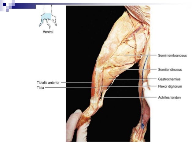
Cat Dissection (Head and Shoulder Muscles) Diagram | Quizlet Upgrade to remove ads Only $35.99/year Cat Dissection (Head and Shoulder Muscles) STUDY Learn Flashcards Write Spell Test PLAY Match Gravity Created by krkoehler2020 Terms in this set (8) Masseter Facial muscle for chewing parotid duct Moves secreted Saliva from parotid gland to the mouth mandible Chewing and mouth movement inferior facial vein Cat Muscle Diagram Anatomy - Free PDF File Sharing CAT DISSECTION A LABORATORY GUIDE - District Collaboration ... underlying muscle layer. 2.Continue cutting longitudinally along the midline ... The cat anatomy is similar to,but not identical to,the human. There are no seminal vesi- [Filename: Cat Dissection Guide.pdf] - Read File Online - Report Abuse Labeled Muscle Diagram - TeacherWeb Cat Leg Anatomy with Diagram - Bones, Muscles, and Nerves ... The most important muscles from the cat thigh are the biceps femoris, semitendinosus, semimembranosus, and abductor. Again, the leg region of a cat consists of both extensor and flexor muscles. Most of these extensor and flexor muscles supply to the pes of the cat. The branches of the axillary artery supply to the front leg of a cat. Cat Muscles | Human Anatomy - Quizizz To play this quiz, please finish editing it. INSTRUCTOR-LED SESSION. Start a live quiz. SUPER. Classic. Students progress at their own pace and you see a leaderboard and live results. Instructor-paced BETA. Control the pace so everyone advances through each question together. ASYNCHRONOUS LEARNING.
Cat dissection lab_labeled_images
20 CFR Appendix 1 to Subpart P of Part 404 - Listing of ... The diagram of the left eye illustrates a visual field, as measured with a III4e stimulus, contracted to 30 degrees in two meridians (180 and 225) and to 20 degrees in the remaining six meridians. The visual efficiency percentage of this field is: ((2 × 30) + (6 × 20)) ÷ 5 = 36 percent. B.
Comments
Post a Comment