41 compound microscope ray diagram
Chemistry Project on Analysis of Fertilizers - iCBSE Diagram showing Refraction. DEFINITIONS: 1. Angle of incidence – The angle that the incident ray makes with the normal is known as angle of incidence (“i"). Ð i = Ð AOB. AO – Incident ray. OB – Normal. 2. Angle of refraction – The angle that the refracted ray makes with the normal is known as angle of refraction. Ð r = Ð COQ. OQ ... (PDF) Scanning electron microscopy and x-ray microanalysis ... Scanning electron microscopy and x-ray microanalysis-Goldstein,Newbury.pdf . × Close Log In. Log in with Facebook Log in with Google. or. Email. Password. Remember me on this computer. or reset password. Enter the email address you signed up with and we'll email you a …
› kingdom-animaliaKingdom Animalia - Different Phylum, Classification ... Kingdom Animalia Different Phylum, Classification, Characteristics Overview. The Kingdom Animalia is a large group that consists of eukaryotic, multicellular organisms that are heterotrophic in nature.

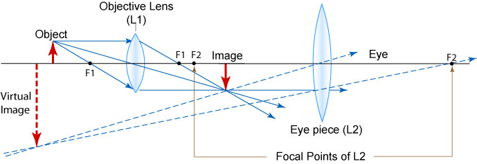
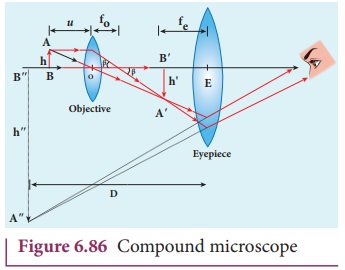
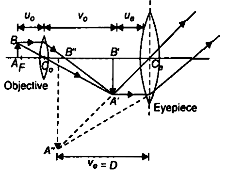
Compound microscope ray diagram
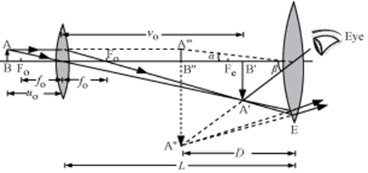
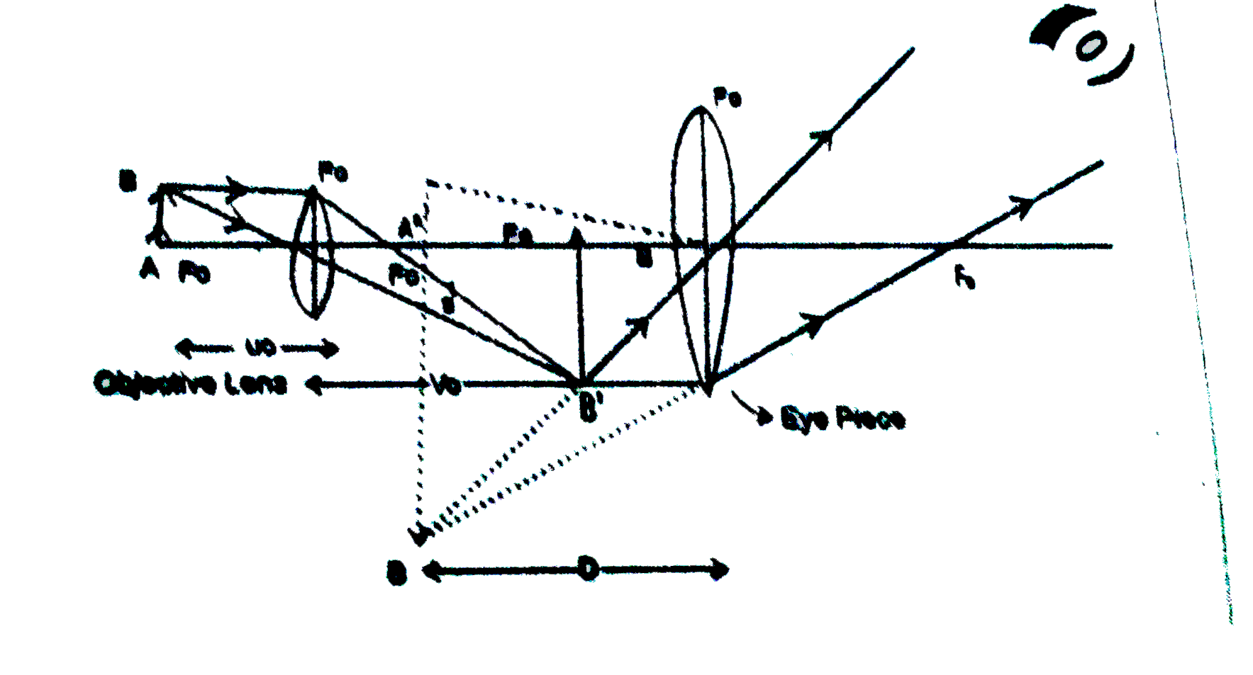
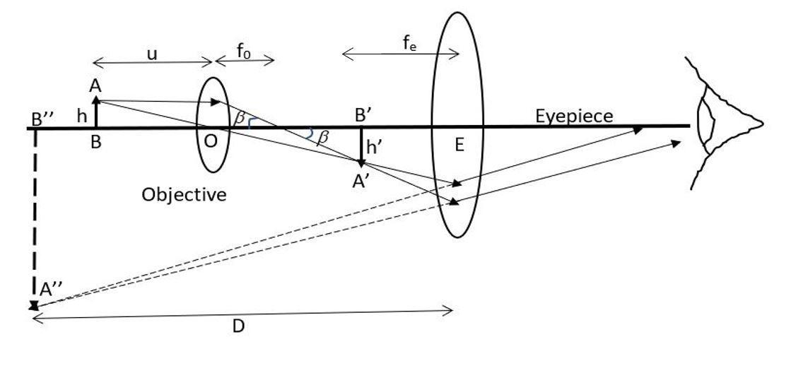
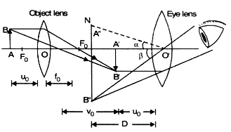
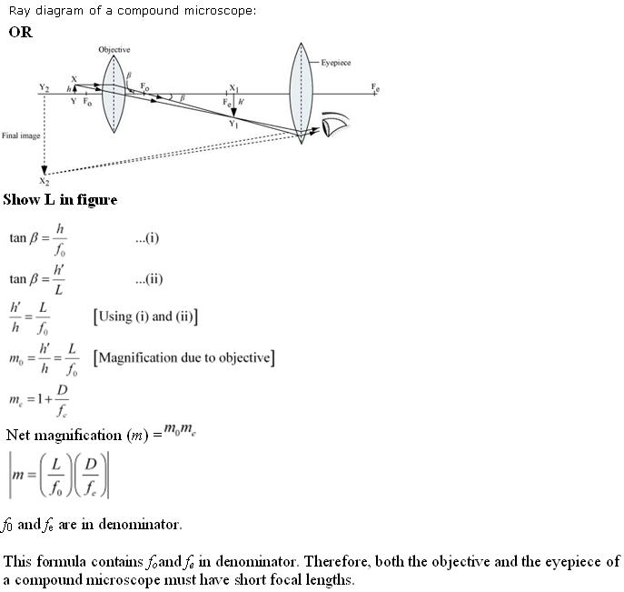
› topics › materials-scienceTransmission Electron Microscopy - an overview ... Transmission electron microscopy (TEM) analysis is conducted to get the actual size of the nanocrystalline cellulose fibers and in some cases the morphology. Nasseri and Mohammadi [99] obtained individual cellulose whiskers with length (L) of 87±28 nm and diameter (d) of 15±3 nm, with an average aspect ratio (L/d) of whiskers obtained was 6±2. en.wikipedia.org › wiki › Electromagnetic_spectrumElectromagnetic spectrum - Wikipedia The electromagnetic spectrum is the range of frequencies (the spectrum) of electromagnetic radiation and their respective wavelengths and photon energies.. The electromagnetic spectrum covers electromagnetic waves with frequencies ranging from below one hertz to above 10 25 hertz, corresponding to wavelengths from thousands of kilometers down to a fraction of the size of an atomic nucleus. › study › bookDraw a ray diagram of a compound microscope. Write the ... Ray diagram of a compound microscope.When the final image is formed at the least distance of distinct vision,For the image formed at infinity, ue = feand By making focal length of the objective small, the magnifying power can be increased.
Compound microscope ray diagram. › technology › microscopemicroscope | Types, Parts, History, Diagram, & Facts | Britannica The most familiar type of microscope is the optical, or light, microscope, in which glass lenses are used to form the image. Optical microscopes can be simple, consisting of a single lens, or compound, consisting of several optical components in line. The hand magnifying glass can magnify about 3 to 20×. Single-lensed simple microscopes can ... › study › bookDraw a ray diagram of a compound microscope. Write the ... Ray diagram of a compound microscope.When the final image is formed at the least distance of distinct vision,For the image formed at infinity, ue = feand By making focal length of the objective small, the magnifying power can be increased. en.wikipedia.org › wiki › Electromagnetic_spectrumElectromagnetic spectrum - Wikipedia The electromagnetic spectrum is the range of frequencies (the spectrum) of electromagnetic radiation and their respective wavelengths and photon energies.. The electromagnetic spectrum covers electromagnetic waves with frequencies ranging from below one hertz to above 10 25 hertz, corresponding to wavelengths from thousands of kilometers down to a fraction of the size of an atomic nucleus. › topics › materials-scienceTransmission Electron Microscopy - an overview ... Transmission electron microscopy (TEM) analysis is conducted to get the actual size of the nanocrystalline cellulose fibers and in some cases the morphology. Nasseri and Mohammadi [99] obtained individual cellulose whiskers with length (L) of 87±28 nm and diameter (d) of 15±3 nm, with an average aspect ratio (L/d) of whiskers obtained was 6±2.
Draw a labelled ray diagram of an image formed by compound ...

Draw a neat and labelled diagram of image formed by a compou ...
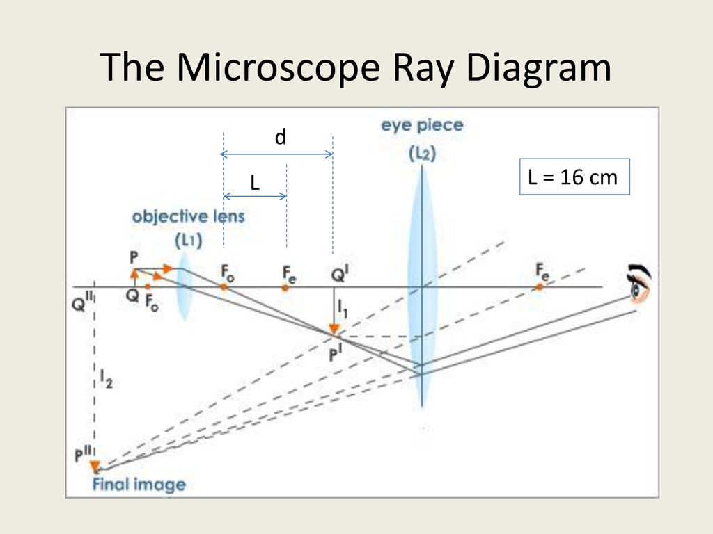
The Compound Microscope - ppt download
Compound Microscope, Ray Diagram Mistakes. | Physics Forums

Draw A Ray Diagram Of A Compound Microscope - Diagram ...
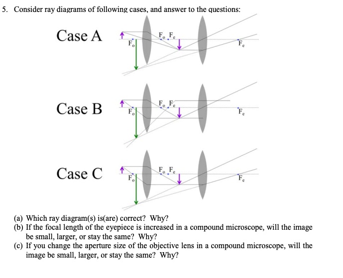
SOLVED:Consider ray diagrams of following cases, and answcr ...

Draw a ray diagram of compound microscope, when final image ...

Leed Lens And Ray Diagram - Electron Microscope Diagram ...

Draw a ray diagram to show formation of an image by a ...

What is a Compound microscope? Applications of Compound ...
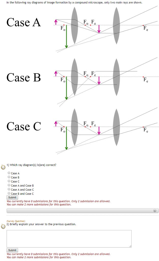
Solved In the following ray diagrams of image formation by a ...
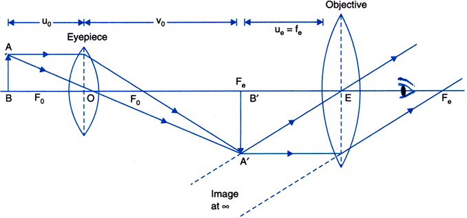
Draw a ray diagram of a compound microscope. Write the ...

optics - Ray diagram of focussing on a compound microscope ...
a) Draw a ray diagram showing the image formation by a ...

Compound Microscope – E-Physics

Optical Instruments: Compound Microscope and its Magnification

compound microscope and telescope off 71% - medpharmres.com

With the help of ray diagram, describe the construction ...

Draw a Ray Diagram Showing the Image Formation by a Compound ...
Draw ray diagram showing the image formation in a compound ...
i) Draw a neat labelled ray diagram of a compound microscope ...
MAGNIFICATION IN MICROSCOPE | cell in life

a)Draw a ray diagram of compound microscope for the final ...
a) Draw a ray diagram for final image formed at distance of disti
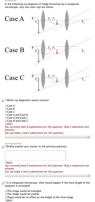
Solved In the following ray diagrams of image formation by a ...

Explain the construction and working of a compound microscope ...
The final image formed by a compound microscope is class 12 ...

Draw a ray diagram of a compound microscope for the final ...
Microscopes | Physics
Simple microscope | Fun Science

Draw the ray diagram of image formation in case of compound ...

CBSE NCERT Notes Class 12 Physics Ray Optics Optical Instruments
Derive the formula for angular magnification of a compound ...

Draw a labelled diagram of an image formed by a compound ...

Show by Ray diagram, the formation of image in case of a ...
Compound microscope - Optical Instruments

(a) Draw a labelled ray diagram showing the formation of a final image by a compound microscope
draw the labelled ray diagram for the formation of image by a ...

A compound microscope has an eyepiece of focal length 10 cm

Draw a neat labelled diagram of a compound microscope and explain
Draw a ray diagram showing image formation in a compound ...
Comments
Post a Comment