39 foot nerve diagram
Sciatic Nerve Diagram High Res Illustrations - Getty Images Browse 26 sciatic nerve diagram stock illustrations and vector graphics available royalty-free, or start a new search to explore more great stock images and vector art. male nervous system, illustration - sciatic nerve diagram stock illustrations. brain and nerves engraving - sciatic nerve diagram stock illustrations. A Complete Guide To The Nerves In Your Feet - Foot Vitals Problems with nerves in the feet are very common. Many times, an injured nerve will cause intense pain and heat felt within the foot. Nerves act as a network, communicating important information from the foot to the brain. Learn more about the various conditions and problems that can affect the nerves in the foot.
Dorsal Foot Diagram Diagram - Quizlet O: Upper lateral surface of fibula, head of fibula, and occasionally the lateral tibial condyle. I: Undersurface of lateral sides of distal end of medial cuneiform and base of metatarsal I. Fibularis longus (function) eversion and plantarflexion of foot. supports arches of foot. Fibularis brevis. O: Lower 2/3 of lateral surface of shaft of fibula.
Foot nerve diagram
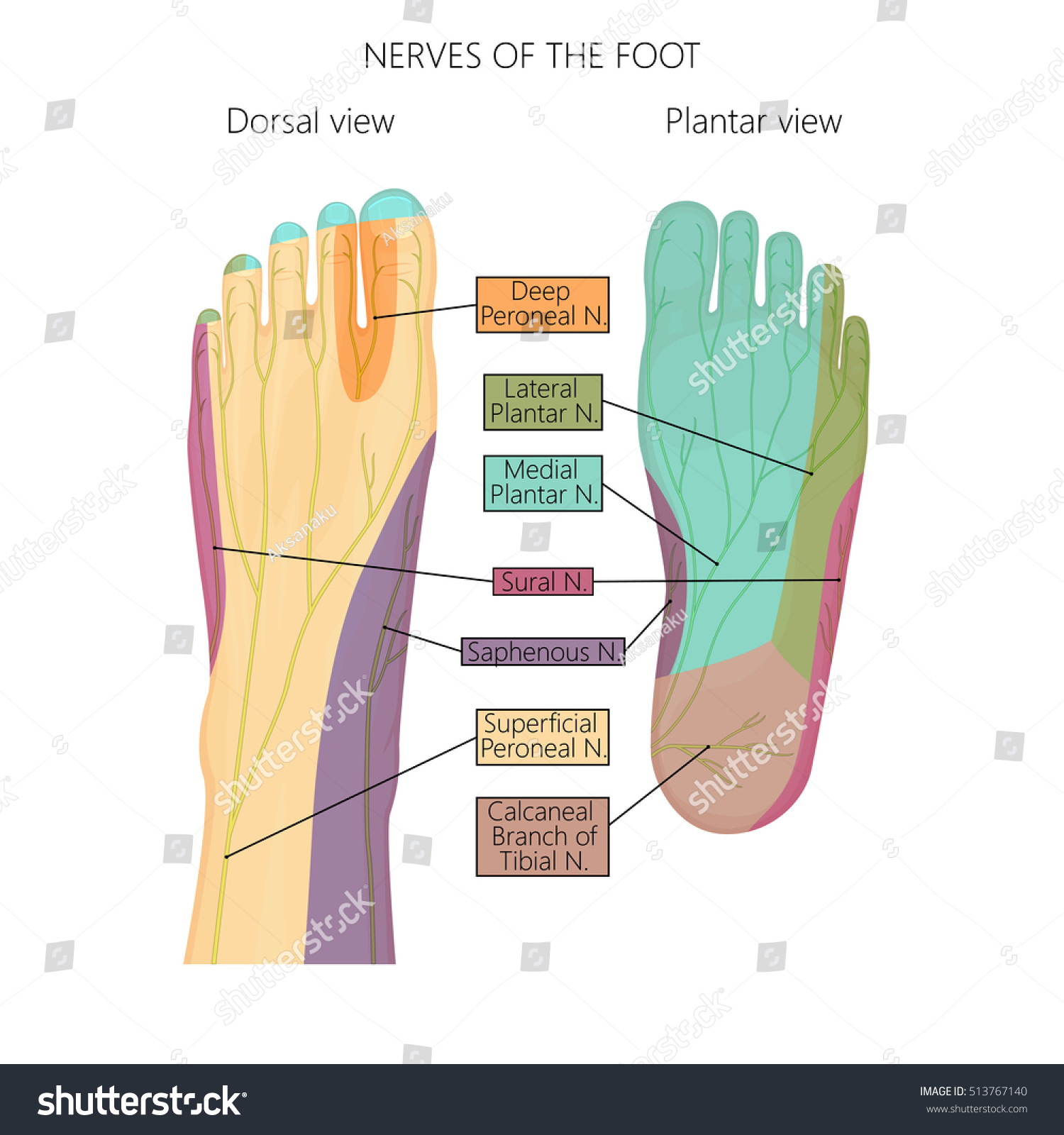
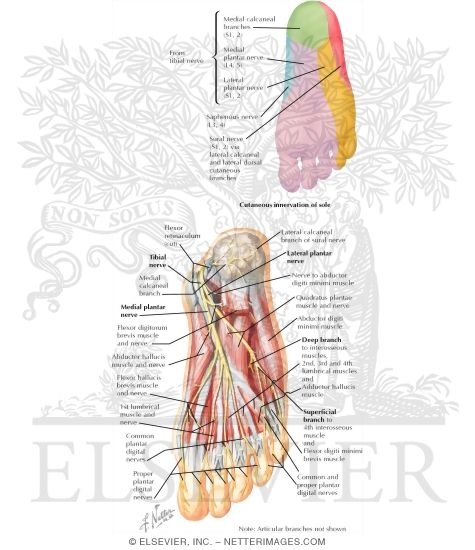
Two diagrams of a foot, one of bones and blood vessels ... Black and white illustration of two diagrams of a foot. The one on the left shows the bones, blood vessels, and nerves of the foot while the one on the right shows the foot with skin on. Alternate Text. Illustration of two diagrams of a foot. Nerves of the foot stock vector. Illustration of ... Illustration about Vector illustration diagram of the nerves and cutaneous innervation of the human foot with palmar and dorsal view. Used transparency. Illustration of neuropathy, deep, dorsal - 80393089 Foot Medical Diagram Photos and Premium High Res Pictures ... the crural nerve - foot medical diagram stock illustrations. old engraved illustration of various kinds of dislocation of bones - foot medical diagram stock pictures, royalty-free photos & images. anatomy of the vascular system engraving antique illustration, published 1851 - foot medical diagram stock illustrations.
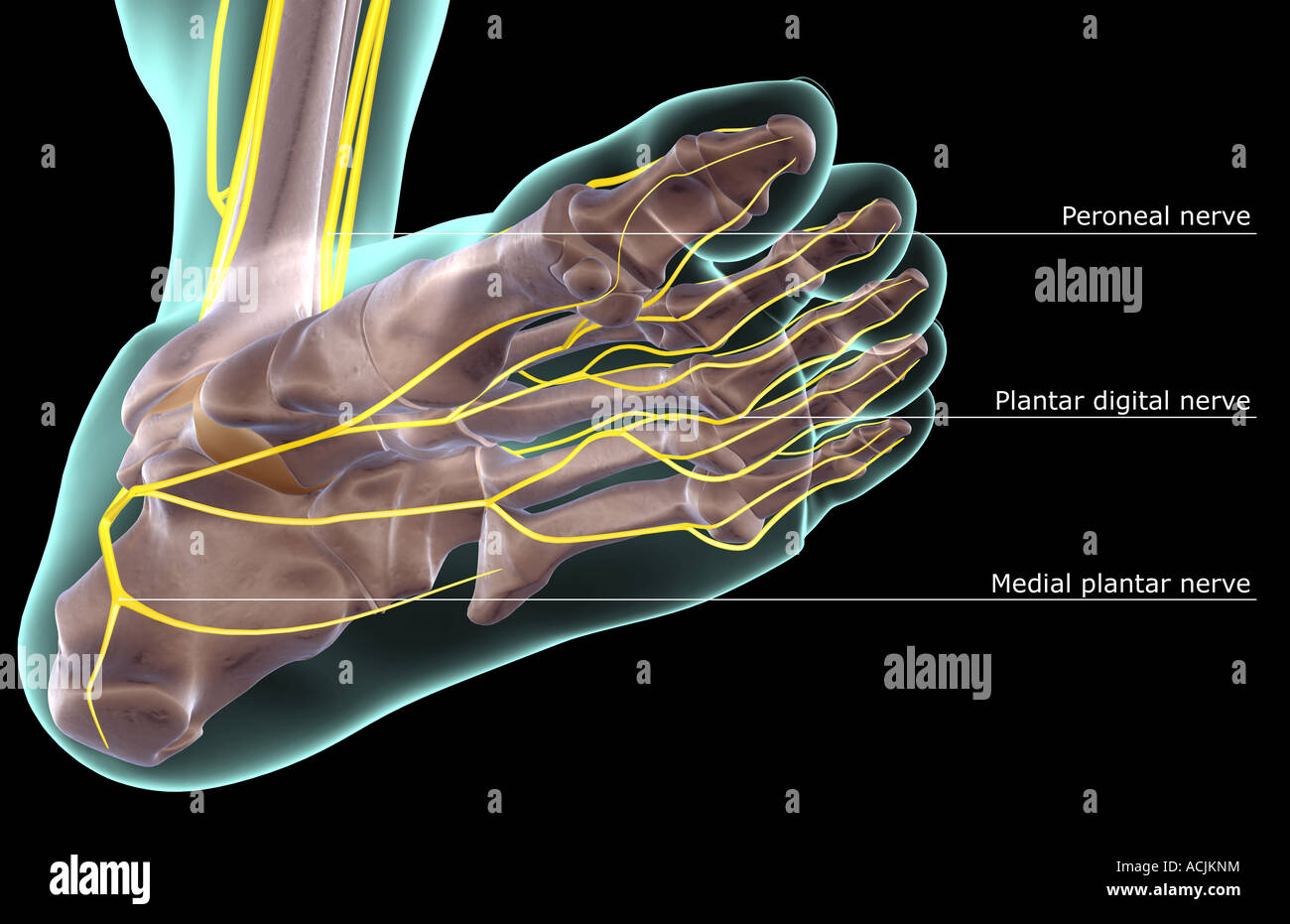
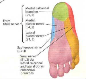
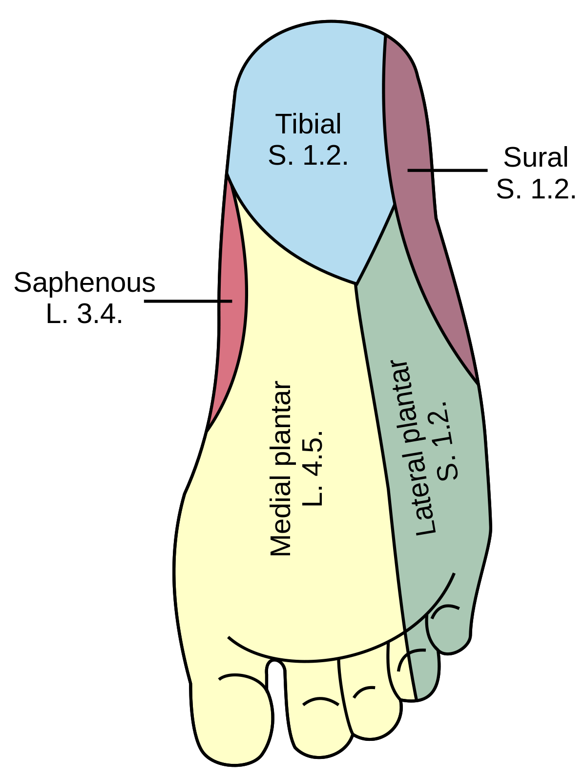
Foot nerve diagram. Nerves of Foot - Earth's Lab Aug 08, 2018 · The deep fibular nerve is parallel and lateral to the tendon of the extensor hallucis longus muscle and goes inside the dorsal aspect of the foot on the lateral aspect of the dorsalis pedis artery. The nerve produces a lateral branch just distal towards the ankle joint, which stimulates the extensor digitorum brevis from its deep surface. Nerves of the Leg and Foot | Interactive Anatomy Guide Jul 03, 2018 · The nerves of the foot help move the body and keep balance both while it’s moving and at rest. All of these nerves extend as branches of nerves in the leg that pass through the ankle and into the foot. The sural nerve branches from the tibial and common fibular nerves and is responsible for feeling on the outside of the foot and the small toe. What Are the Nerves of the Foot? (with pictures) The medial plantar nerve is found on the big-toe side of the foot. It supplies the muscles that flex or curl the toes as well as those that abduct and adduct, or fan out and bring together, the toes. It also innervates the skin on this half of the sole of the foot, including the plantar surfaces of the first three and a half toes. Are Nerve Problems Causing Your Foot Pain? - Verywell Health Four common nerve problems can cause foot pain: Morton's neuroma, tarsal tunnel syndrome, diabetic peripheral neuropathy, and a pinched nerve. You'll probably know when trouble strikes. Nerve problems often trigger burning or shooting pain. And the sensation can be so intense that it can rouse you from a deep sleep.
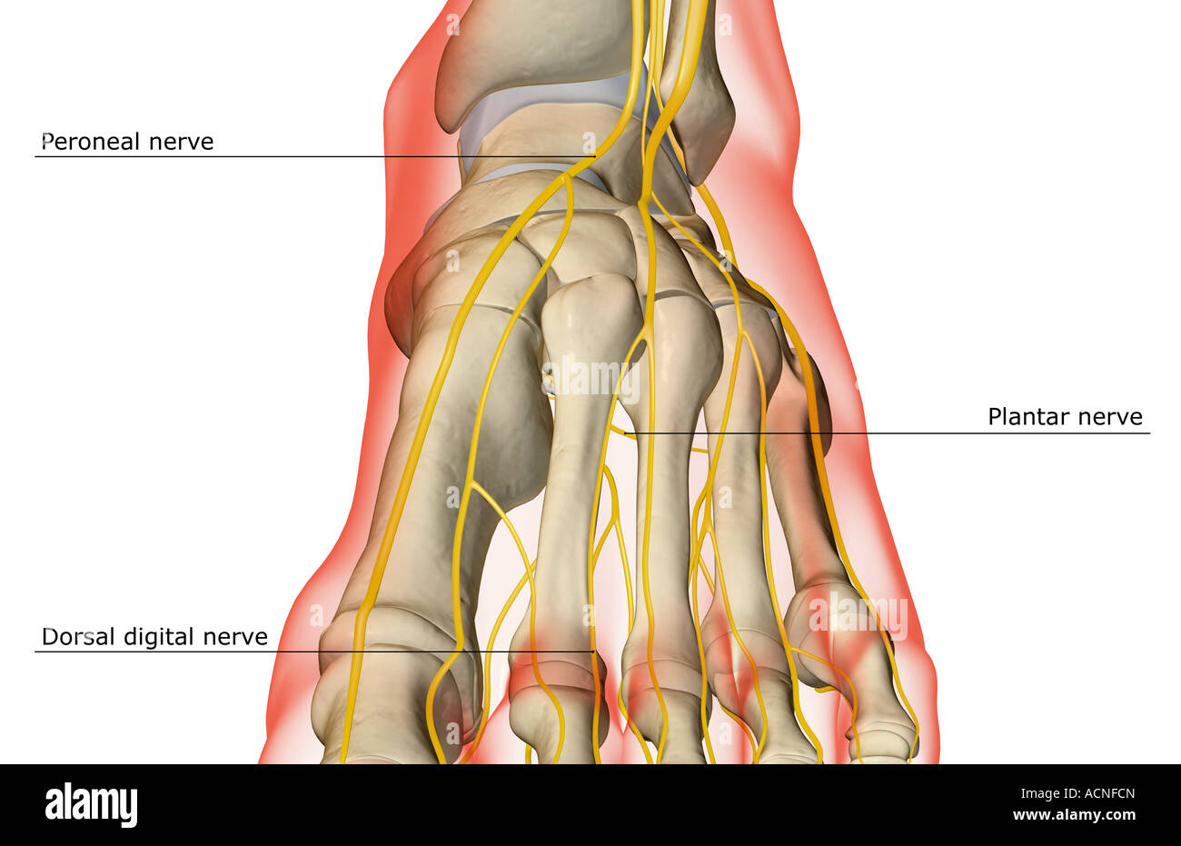
Foot Vessels Anatomy, Function & Diagram | Body Maps The dorsal digital nerves of the foot branch throughout the body of the foot and down through each toe. They are themselves branches of the larger intermediate dorsal cutaneous nerve, medial dorsal... Diagram Of Sciatic Nerve Pathway - Wiring Diagrams The sciatic nerve (also called ischiadic nerve, ischiatic nerve) is a large nerve in humans and other animals. It begins in the lower back and runs through the buttock and down the lower limb. It is the longest and widest single nerve in the human body, going from the top of the leg to the foot on the posterior aspect. Sciatic Nerve Anatomy - Spine-health The sciatic nerve is the largest and longest nerve in the human body, originating at the base of the spine and running along the back of each leg into the foot. 1, 2 At its thickest point, it is about as wide as an adult thumb. The sciatic nerve is formed in the lower spine by the combination of motor and sensory fibers from spinal nerves L4 to S3. How to Alleviate Foot Nerve Pain (6 simple tips) Why Foot Nerve Pain Happens. There are many potential reasons for foot nerve pain, including an injury, a disease such as diabetes, chemotherapy, certain lower back conditions, side effects of medications, and more.1 But the exact mechanism that causes the pain is still unclear.
human foot nerve diagram - MedHelp A high arch is the opposite of a flat foot, and somewhat less common. The term pes cavus encompasses a broad spectrum of foot deformities. However, a pain management doctor gave me a diagram of the L5 and S1 nerve, showing they split off. He also said in spinal fusion, this is more often than not the outcome. Nerves of the Foot - Foot & Ankle - Orthobullets 3%. (71/2762) 3. It is the terminal branch of the superficial peroneal nerve; injury leads to reduced sensation over medial aspect of great toe. 83%. (2287/2762) 4. It is the terminal branch of the deep peroneal nerve; injury leads to first interphylangeal joint flexion weakness. 3%. Normal Anatomy and Compression Areas of Nerves of the Foot ... normal anatomy and common variants of the nerves of the foot and ankle, with use of dissected specimens and correlative US and MR imaging findings. In addition, the authors illustrate proper probe positioning, which is essential for visualizing the nerves at US. The authors' discussion focuses on the superficial and deep Nerves Of The Leg & Foot - Everything You Need To Know ... Dr. Ebraheim's educational animated video describes the nerves of the lower leg in a very easy and simple animation.Lateral cutaneous nerve of the calfSural ...
Nerves Of The Foot High Resolution Stock Photography and ...
Nerves of the Foot - SmartDraw Nerves of the Foot. Create healthcare diagrams like this example called Nerves of the Foot in minutes with SmartDraw. SmartDraw includes 1000s of professional healthcare and anatomy chart templates that you can modify and make your own.

Schematic diagram of anatomical variations of the plantar ...
Sciatic Nerve Anatomy, Location & Diagram | Body Maps Sciatic nerve. The sciatic nerve is the dominant nerve that innervates the lower back and the lower extremities. It travels from the lower spine, through the pelvis, and down each leg. It is the ...

Foot nerves Images, Stock Photos & Vectors | Shutterstock
Nerve Pain In Foot: Causes, Symptoms & Diagnosis At the base of the spine, five nerves (L4, 5, S1, 2 &3) exit the spine and join together to form the sciatic nerve, the largest nerve in the body. The sciatic nerve travels down through the buttock and the back of the leg, branching out as it spreads further down through the leg and into the foot.

foot | Description, Drawings, Bones, & Facts | Britannica
Diagram Of Sciatic Nerve Pathway - schematron.org Diagram Of Sciatic Nerve Pathway; Diagram Of Sciatic Nerve Pathway. The sciatic nerve is the dominant nerve that innervates the lower back and the lower extremities. It travels from the lower spine, through the pelvis, and down each leg. When something injures or puts pressure on the sciatic nerve, it can cause pain in the lower back that ...

Normal Anatomy and Compression Areas of Nerves of the Foot ...
nerves of the leg diagram - ModernHeal.com This image is titled nerves of the leg diagram and is attached to our article about Leg Nerves and Reflex Motion in Feet.. Be sure to visit the guide for more context and information about nerves of the leg diagram, or read some of our other Health & Anatomy posts!

The Tibial Nerve - Course - Motor - Sensory - TeachMeAnatomy
Nerve pain in the foot: Symptoms, causes, treatment, and more Nerve pain in the foot tends to result from a compressed nerve or diabetes. A range of health issues may be at play, and they tend to cause similar symptoms. For this reason, receiving a diagnosis ...
Vector Illustration Diagram Nerves Cutaneous Innervation ...
Anatomy of the Foot and Ankle - OrthoPaedia Sural Nerve. The fourth nerve of the foot is another branch of the tibial nerve, known as the sural nerve (Figure 17). This nerve runs from slightly below the knee to the lateral aspect of the foot. It becomes a very superficial nerve at the level of the posterolateral ankle and continues distally to provide sensation to the outside of the foot.
Lower limb arteries and nerves: Anatomy, branches | Kenhub
Spinal Nerve Chart - Miller Chiropractic Clinic On the chart below you will see 4 Columns (Vertebral Level, Nerve Root, Innervation, and Possible Symptoms). Under 'Vertebral Level': C1-C7 is the NECK, ; T1-T12 is the UPPER BACK/rib cage area, and ; L1-L5 is the LOWER BACK.; Simply line up the "Vertebral Level" with the "Possible Symptoms" and you will see some surprising connections of symptoms that relate to your spine.

Human foot nerves Stock Photos and Images | agefotostock
Foot Condition By Area | Top of Foot | Foot Pain Diagram ... Match the corresponding numbers on the foot diagram below for a list of conditions that may be causing your foot and ankle pain. This is meant for educational purposes only. If you're having a problem with your foot or ankle, visit a podiatrist - a foot and ankle specialist! Top (Dorsal) View of Foot & Ankle Number 1 and 2:
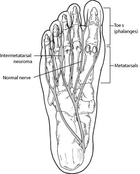
Morton's Neuroma – Symptoms of Morton's Neuroma | Foot Health ...
Anatomy of the foot | Structural diagram of foot | Patient The foot contains a lot of moving parts - 26 bones, 33 joints and over 100 ligaments. The foot is divided into three sections - the forefoot, the midfoot and the hindfoot. The forefoot. This consists of five long metatarsal bones and five shorter bones that form the toes (phalanges).
The Diagnostic Anatomy of the Sciatic Nerve | Neupsy Key
Foot Pain Diagram - Why Does My Foot Hurt? The first foot pain diagram looks at the front and top of the foot, the second foot pain identifier looks underneath and at the back of the foot. Front Foot Pain Identifier This foot pain diagram shows common problems that cause pain on top of the foot at the front. A. Sinus Tarsi Syndrome

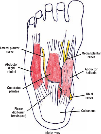
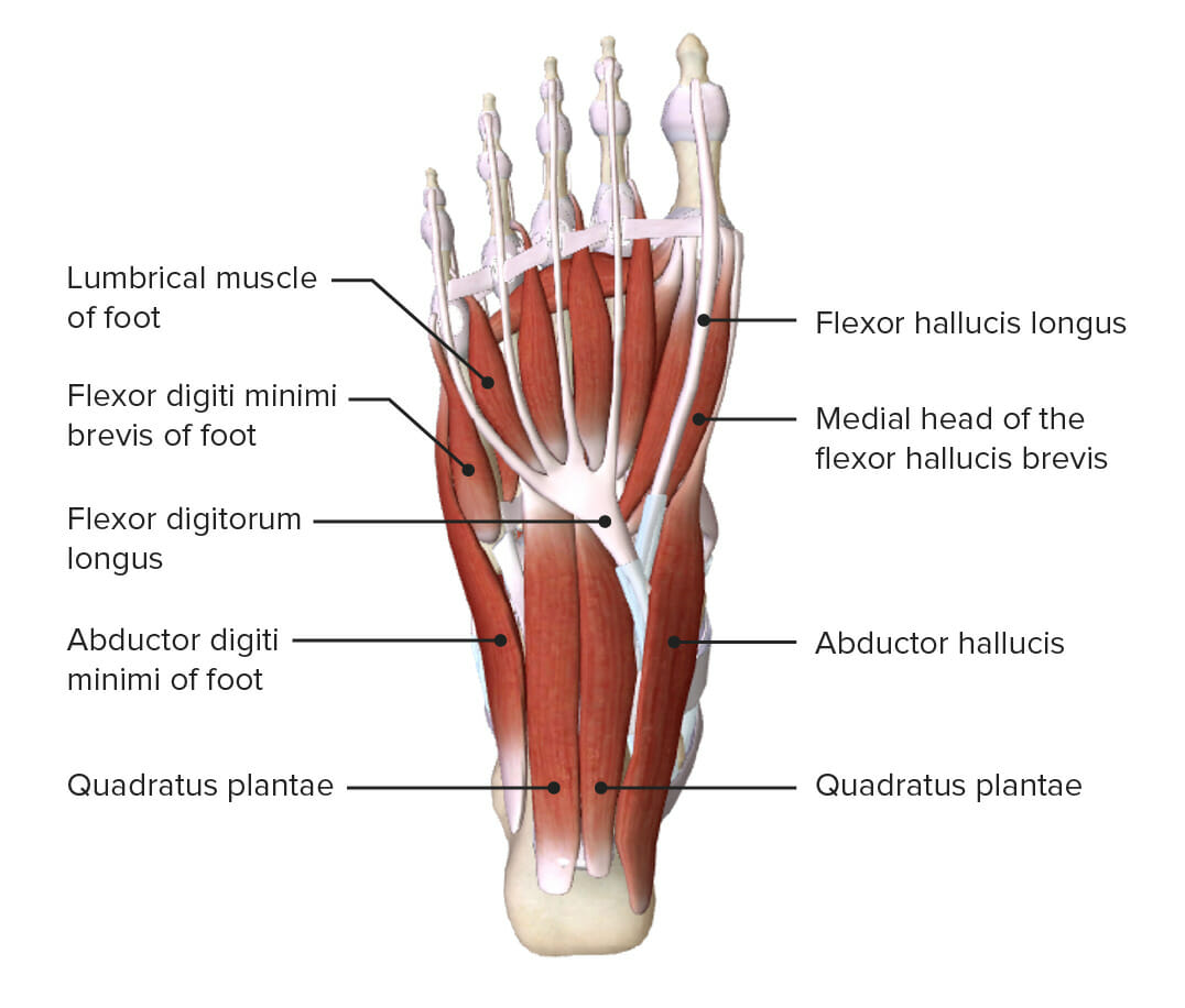
7 The structures of the plantar aspect of the right foot. The ...
Foot Medical Diagram Photos and Premium High Res Pictures ... the crural nerve - foot medical diagram stock illustrations. old engraved illustration of various kinds of dislocation of bones - foot medical diagram stock pictures, royalty-free photos & images. anatomy of the vascular system engraving antique illustration, published 1851 - foot medical diagram stock illustrations.

Central Muscles And Bones Of The Foot Sole Labeled Human ...
Nerves of the foot stock vector. Illustration of ... Illustration about Vector illustration diagram of the nerves and cutaneous innervation of the human foot with palmar and dorsal view. Used transparency. Illustration of neuropathy, deep, dorsal - 80393089

The Tibial Nerve - Course - Motor - Sensory - TeachMeAnatomy
Two diagrams of a foot, one of bones and blood vessels ... Black and white illustration of two diagrams of a foot. The one on the left shows the bones, blood vessels, and nerves of the foot while the one on the right shows the foot with skin on. Alternate Text. Illustration of two diagrams of a foot.

Anatomy of the foot | Osmosis

Medial Muscles And Bones Of The Foot Sole Labeled Human ...

Dorsal digital nerves of foot - Wikipedia

Anatomical dissection of the cutaneous nerves of the foot and ...
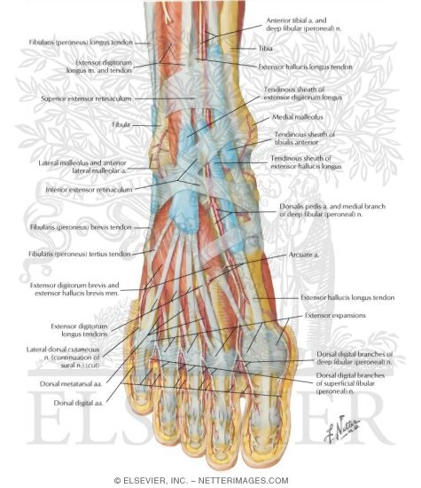
Muscles, Arteries, and Nerves of Front of Ankle and Dorsum of ...

Foot Human anatomy Nerve Muscle Muscular system, chinese arch ...

Nerves of the foot-02 stock vector. Illustration of mortons ...

Normal Anatomy and Compression Areas of Nerves of the Foot ...

HealthCrib su Twitter: "THE FOOT: nerves of the foot that ...

Foot nerves Images, Stock Photos & Vectors | Shutterstock
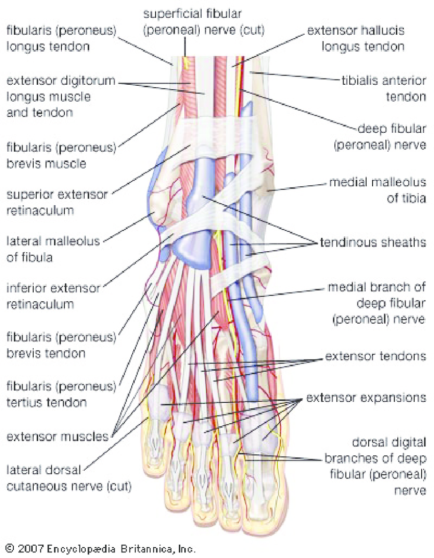
Nerves of foot. (adapted from Encyclopedia Britanica 2007 ...

Compression Neuropathy - Lincoln Park | Lakeview Chicago, IL ...

Foot and Ankle | Musculoskeletal Key

Nerves of the Foot | ClipArt ETC | Anatomy organs, Human ...

Nerve and Tendon Lacerations About the Foot and Ankle | Nerve ...

Thumb Human leg Nerve Knee Muscle, Human Bein, angle, hand ...

Tibial Nerve - an overview | ScienceDirect Topics

Ankle Block - Landmarks and Nerve Stimulator Technique ...

The leg, ankle, and foot - Knowledge @ AMBOSS
Foot: Anatomy | Concise Medical Knowledge
Medial Plantar Nerve - Physiopedia
Plantar nerve - Wikipedia

Foot anatomy, Nerve anatomy, Ankle anatomy
The nerves of the foot Stock Photo - Alamy
Plantar Nerves and Foot Sensory
Comments
Post a Comment