42 pseudostratified columnar epithelium diagram
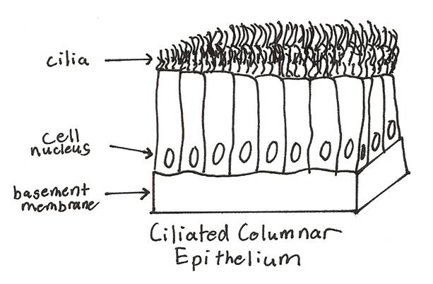
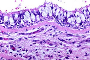
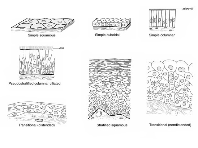
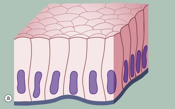
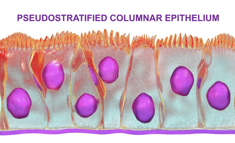
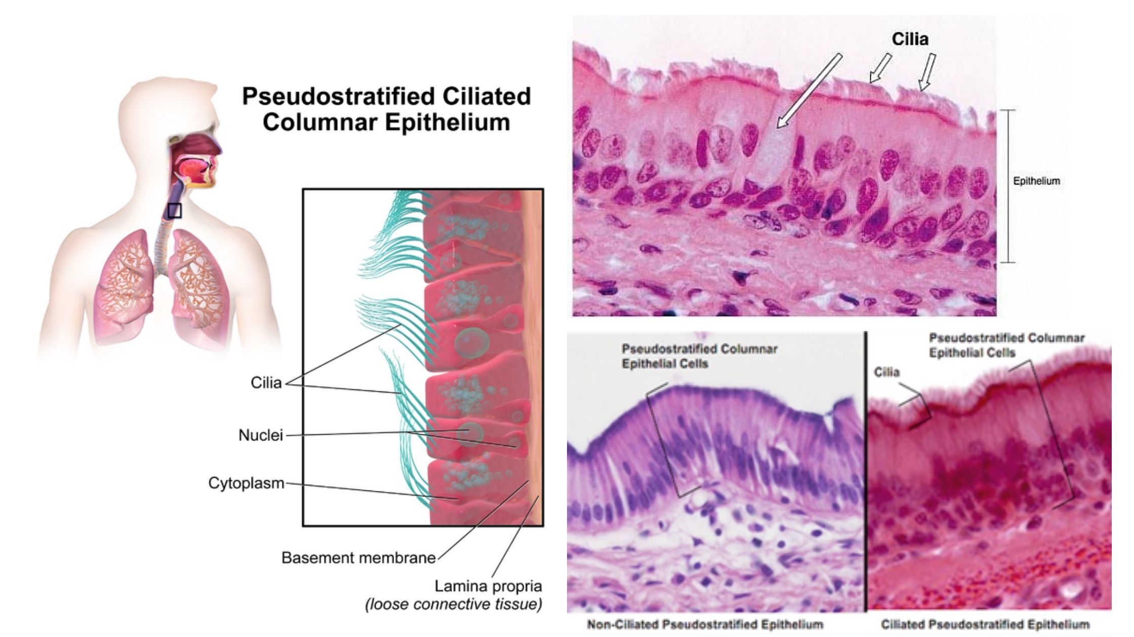
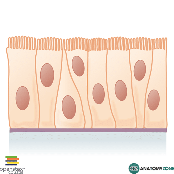
Unlike the epithelium of the skin, a pseudostratified ciliated columnar epithelium appears to have multiple layers, but is actually only comprised of a single sheet of cells. The positioning of the nuclei within the individual columnar cells causes this illusion. These structures, which are easily identifiable with the help of a microscope, are ... Transitional epithelium also known as urothelium is a type of stratified epithelium. Transitional epithelium is a type of tissue that changes shape in response to stretching (stretchable epithelium). The transitional epithelium usually appear cuboidal when relaxed and squamous when stretched. This tissue consists of multiple layers of epithelial cells which can contract and …
Feb 24, 2016 - pseudostratified ciliated columnar epithelium diagram - Google Search
Pseudostratified columnar epithelium diagram
Pseudostratified Columnar Epithelium Definition. Pseudostratified columnar epithelia are tissues formed by a single layer of cells that give the appearance of being made from multiple layers, especially when seen in cross section. The nuclei of these epithelial cells are at different levels leading to the illusion of being stratified. Pseudostratified Columnar Epithelium (Figure 4.3d) The cells of pseudostratified (soo-do-strat′˘-f ¯ı ıd) columnar epithelium are varied in height. All of its cells rest on the basement membrane, but only the tall cells reach the apical surface of the epithelium. The short cells are undifferentiated Pseudostratified Columnar Epithelium •Secretion of mucus. Identify the structure indicated. Stratified Cuboidal Epithelium Basement Membrane. Identify the tissue type and a location where it is found. Stratified Cuboidal Epithelium •Largest ducts of sweat, mammary and salivary glands.
Pseudostratified columnar epithelium diagram. The pseudostratified columnar epithelium is a type of epithelium consisting of a single layer of cells that gives the appearance of being multiple layers because the nuclei of the cells are present at different levels. This epithelium is histologically a simple epithelium even though in a crosssection, it might appear as a stratified epithelium. Pseudostratified Columnar Epithelium. This diagram represents pseudostratified columnar epithelium.This is a special type of simple epithelium called pseudostratified epithelium as it resembles stratified epithelium due to the positioning of the cellular nuclei, but is comprised of only a single layer of cells. A. Simple columnar epithelium. Slide 29 (small intestine) View Virtual Slide Slide 176 40x (colon, H&E) View Virtual Slide Remember that epithelia line or cover surfaces. In slide 29 and slide 176, this type of epithelium lines the luminal (mucosal) surface of the small and large intestines, respectively. Refer to the diagram at the end of this chapter for the tissue orientation and … Pseudostratified columnar epithelium is a type of epithelium that appears to be stratified but instead consists of a single layer of irregularly shaped and differently sized columnar cells. In pseudostratified epithelium, nuclei of neighboring cells appear at different levels rather than clustered in the basal end. The arrangement gives the ...
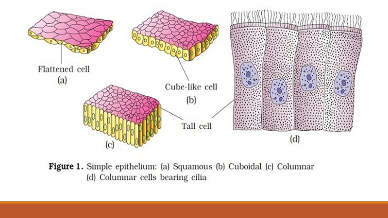
Simple epithelium ; Pseudostratified columnar, Location: trachea and most of the upper respiratory tract (ciliated cells) Function: secretes mucus which is moved ...Pseudostratified columnar: Location: trachea a...Function and classes: Function: absorption and ...Simple columnar: Location: bronchi, uterine tu...Simple cuboidal: Location: secretory ducts of s... Pseudostratified Columnar: Respiratory passage and ducts of many glands: Similar to columnar epithelium but all the cells are not of similar height: Protection, secretion and movement of mucous: Transitional epithelia or urothelium: Urinary bladder, urethra, ureter: Stratified epithelium, which can contract or expand as per the requirement. Sep 27, 2020 · The simple columnar epithelium is a type of epithelium that is formed of a single layer of long, elongated cells mostly in areas where absorption and secretion are the main functions. Like cuboidal epithelium, the cells in the columnar epithelium are also modified to suit the function and structure of the organ better. A. Simple columnar epithelium. Slide 29 (small intestine) View Virtual Slide Slide 176 40x (colon, H&E) View Virtual Slide Remember that epithelia line or cover surfaces. In slide 29 and slide 176, this type of epithelium lines the luminal (mucosal) surface of the small and large intestines, respectively. Refer to the diagram at the end of this chapter for the tissue orientation and …
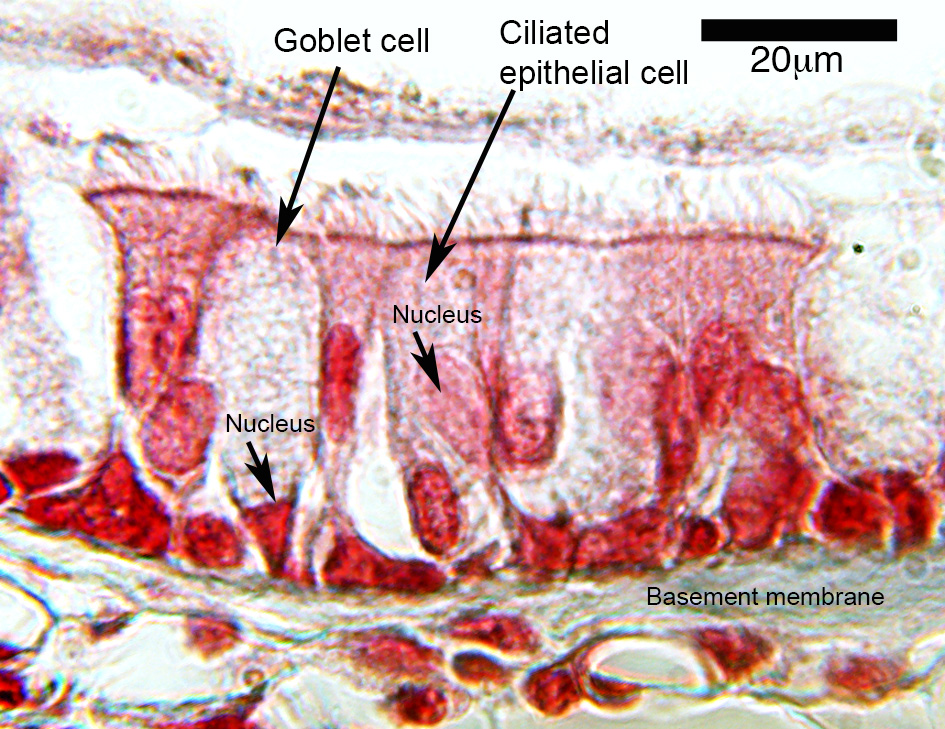
Pseudostratified columnar. All cells rest on a basement membrane. Single layer, but some cells are shorter than others giving a false (pseudo) impression of stratification. Location: Respiratory tract, where it is ciliated and known as pseudostratified ciliated columnar epithelium. Functions in absorption or secretion Diagrammatic Illustration Of The Pseudostratified Columnar Epithelium (Source: dartmouth.edu) The diagram above illustrates a pseudostratified epithelium. In terms of shape, the cells are variously shaped but most of them are taller than wide (as implied by their name, columnar) and are shown to appear like miniature pillars. Pseudostratified Columnar Epithelium is a single layer of ciliated, irregularly shaped cells containing many goblet cells. In usual slides the boundaries between epithelial cells are often not clearly seen but because of the shape and spacing of the nuclei, the epithelium can be identified. The Pseudostratified Columnar Epithelium lining the ... This type of epithelium is adapted for secretion and/or absorption, and can also be protective. Simple secretory columnar epithelium lines the stomach and uterine schematron.org simple columnar epithelium that lines the intestine also contains a few goblet cells. Simple Columnar Epithelium: A Labeled Diagram and Functions Epithelium is a tissue ...
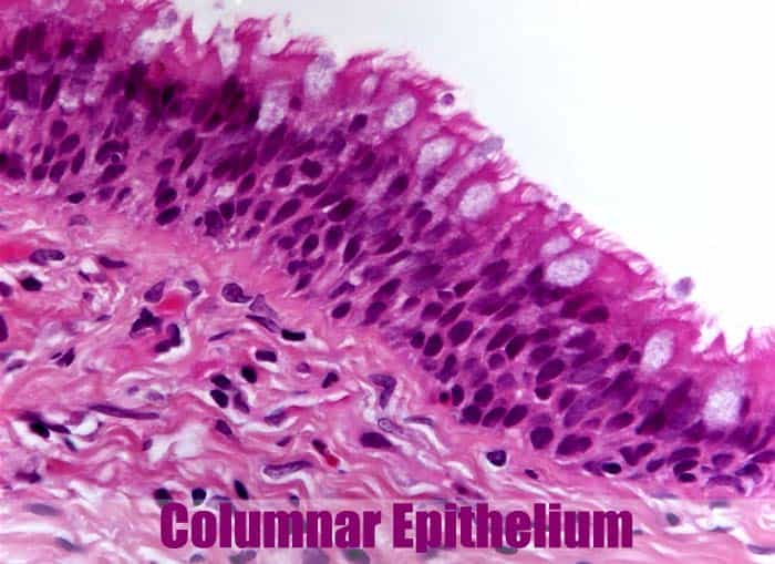
Ciliated Columnar Epithelium
Label the following Tissue in your drawing Diagram Magnification: Basement membrane Principal cells Epididymis Pseudostratified columnar epithelium Basal cell Spermatozoa Tissue Label the following in your drawing Diagram Magnification: Seminal vesicles Pseudostratified columnar epithelium Lumen Basal cells 1. Compare and contrast the functions ...
Pseudostratified Columnar Epithelium - Definition & Function
Holocrine is a term used to classify the mode of secretion in exocrine glands in the study of histology.Holocrine secretions are produced in the cytoplasm of the cell and released by the rupture of the plasma membrane, which destroys the cell and results in the secretion of the product into the lumen.. Holocrine gland secretion is the most damaging (to the cell itself and …
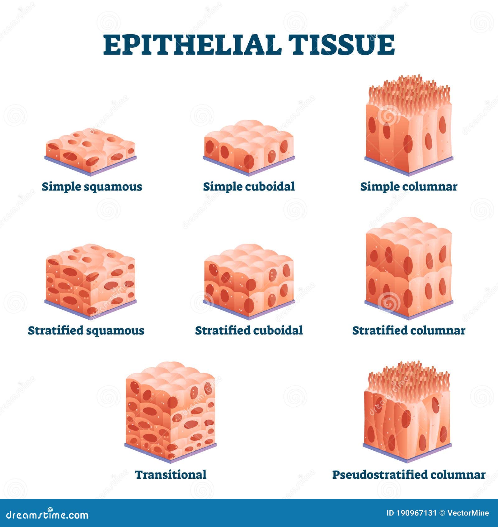
Types of epithelial tissue: simple, compound and specialized ...
Label the parts of the pseudostratified columnar epithelium. Learn with flashcards, games, and more — for free.

0614 Simple Cuboidal Epithelium Medical Images For Powerpoint ...
Observation: Simple Columnar Epithelium Simple columnar epithelium is a single layer of tall, cylindrical cells, each with a nucleus near the base. This tissue, which lines the digestive tract from the stomach to the anus, protects, secretes, and allows absorption of nutrients. 1. 2. 3. Study a model or diagram of simple columnar epithelium ...

File:Pseudostratified columnar epithelium drawing.png ...
Pseudostratified columnar epithelium is tissue composed of a single layer of columnar cells that line the space of an organ cavity or vessel. It is particularly useful in the secretion and ...
Solved Vlew previous atten Simple Simple squamous | Chegg.com
Pseudostratified Ciliated Epithelium. Pseudostratified ciliated columnar epithelia are tissues that are made up of only one layer of cells but appear to be made up of numerous layers when viewed in cross-section. The nuclei of epithelial cells are at extremely different levels, giving the appearance of stratification.

psuedostratified columnar epithelium tissue | Human anatomy ...
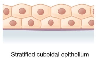
Structure of the transitional epithelium. Transitional epithelium is an epithelial tissue which in a relaxed state appears as a stratified cuboidal epithelium.; The cells in the transitional epithelium are pear-shaped or round, but as tissue is stretched, cells become flattened, giving the appearance of stratified squamous epithelium.; The cells in the basal layer appear cuboidal or columnar ...
Pseudostratified Columnar Epithelium | Histology, Anatomy & Types
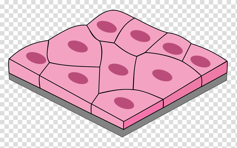
Jul 21, 2021 · The simple squamous epithelium is different from other types of epithelial tissue such as simple cuboidal, simple columnar, and stratified squamous epithelium in that it is only made of one layer ...

Pseudostratified Columnar Epithelium Diagram | Quizlet
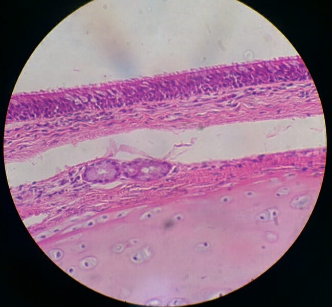
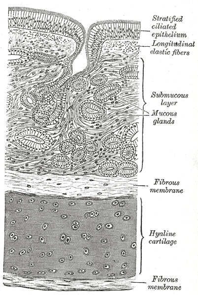
The epithelium is tall columnar pseudostratified with cilia and goblet cells. The supporting lamina propria underneath the epithelium contains elastin, that plays a role in the elastic recoil of the trachea during inspiration and expiration, together with blood vessels that warm the air.
Epithelial Tissue - Definition, types, functions, examples
Start studying Pseudostratified Columnar Epithelium. Learn vocabulary, terms, and more with flashcards, games, and other study tools.
Simple epithelium: Location, function, structure | Kenhub
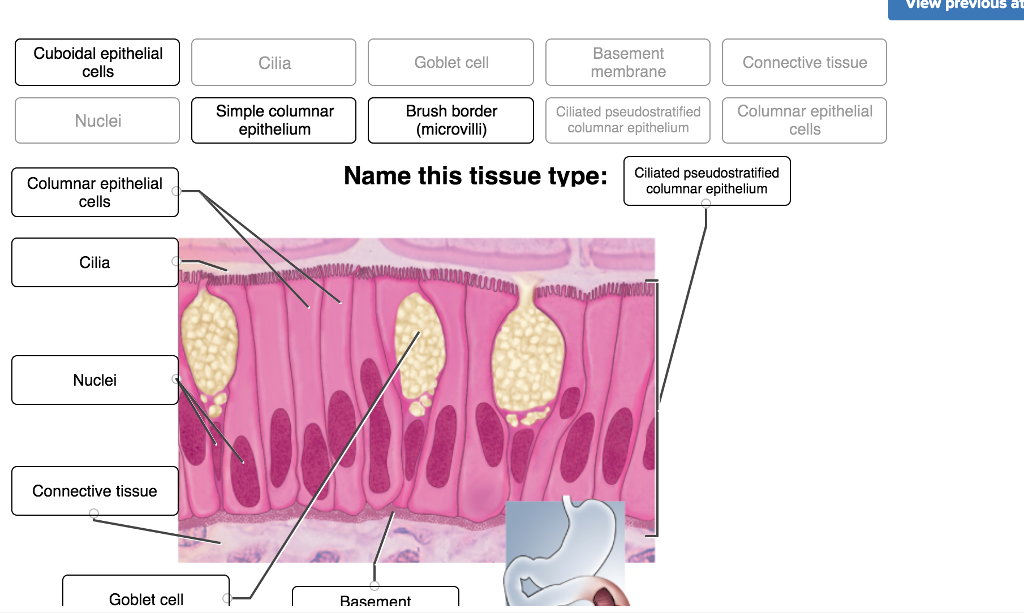
The nasopharynx is primarily lined by two types of epithelia, with the stratified squamous epithelium comprising around 60% of its inner walls [5].The nasopharynx is also the only section of the pharynx to have pseudostratified columnar respiratory epithelium [6], the specialized epithelium (ciliated and containing goblet cells) for the respiratory tract [7].

Pseudostratified Columnar Epithelium Function & Location ...
Diagram of a pseudostratified columnar epithelium. A pseudostratified columnar epithelium is a special type of simple epithelium that consists of a falsely-stratified single layer of epithelial cells resting on a basement membrane. Many, if not most, of the cells, are columnar. The single layer of cells appears stratified because the cells have ...
Ciliated pseudostratified columnar epithelium of the trachea ...
Sep 28, 2020 · Stratified columnar epithelium definition. The stratified columnar epithelium has multiple layers of cells in which the apical layer is made up of columnar cells while the deeper layer can be either cuboidal or columnar. As in the case of other stratified epithelium, the cells in the deeper layers might be different than the layer on the top.
Medpics - UC San Diego, School of Medicine
Epithelium is one of only 4 types of human body tissues.Like all types, it is formed by cells within an extracellular matrix (ECM). The cells in this tissue are tightly packed within a thin ECM. Forming sheets that cover the internal and external body surfaces (surface epithelium) and secreting organs (glandular epithelium). Functions of epithelial tissue are secretion, protection, absorption ...
Simple squamous epithelium Pseudostratified columnar ...
Pseudostratified ciliated columnar epithelium lines the trachea (windpipe) and larger respiratory passage ways. 22. Skeletal (striated) muscle model Lab-2 29. Skeletal muscle is the most abundant type of muscle tissue found in the vertebrate body, making up at least 40% of its mass. Although it is often activated by reflexes that function in ...

Epithelia Cells Objectives Define Epithelia Cells Identify the
The pseudostratified columnar epithelium (with a variety of stereocilia) is also shown on the epididymis labeled diagram. A circularly arranged smooth muscle fibers that surround the epididymal tubules are also identified in the labeled diagram. There is a clump of spermatozoa present in the lumen of the epididymis.
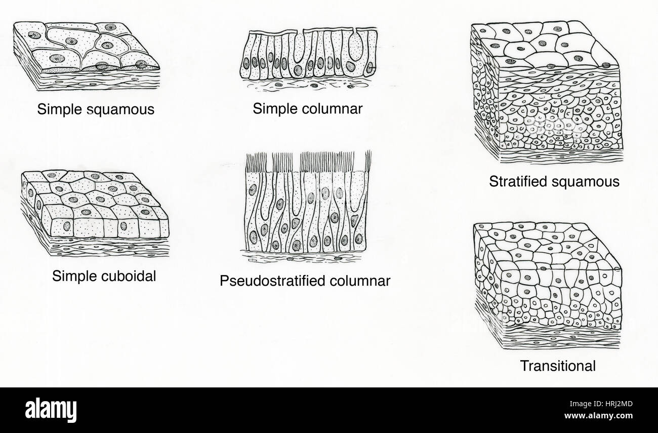
Illustration of Epithelium Types Stock Photo - Alamy
Pseudostratified ciliated epithelium (40X) Human respiratory tract The bar in this image shows the thickness of the layer of ciliated pseudostratified epithelium. The rest of the tissue below the epithelium is mostly connective tissue, but there are some mucous glands (glandular epithelium) just below the surface.

Epithelial Tissues | Biology for Majors II
The epithelial tissue made up of a single layer of epithelial cells of different heights is known as the pseudostratified columnar epithelium.Jul 24, 2021What does Pseudostratified columnar epithelium do?Where can Pseudostratified columnar epithelium be found?

Pseudostratified Epithelium Pseudostratified epithelium Table ...
Pseudostratified Columnar Epithelium •Secretion of mucus. Identify the structure indicated. Stratified Cuboidal Epithelium Basement Membrane. Identify the tissue type and a location where it is found. Stratified Cuboidal Epithelium •Largest ducts of sweat, mammary and salivary glands.
Epithelial tissues | Basicmedical Key
Pseudostratified Columnar Epithelium (Figure 4.3d) The cells of pseudostratified (soo-do-strat′˘-f ¯ı ıd) columnar epithelium are varied in height. All of its cells rest on the basement membrane, but only the tall cells reach the apical surface of the epithelium. The short cells are undifferentiated

Pseudostratified epithelium
Pseudostratified Columnar Epithelium Definition. Pseudostratified columnar epithelia are tissues formed by a single layer of cells that give the appearance of being made from multiple layers, especially when seen in cross section. The nuclei of these epithelial cells are at different levels leading to the illusion of being stratified.

Stratified columnar epithelium - Wikipedia
Pseudostratified Columnar Epithelium Stock Illustration ...

Description

What purpose does the pseudostratified ciliated columnar ...

Simple Columnar Epithelium: A Labeled Diagram and Functions ...

Pseudo stratified columnar. Simple cuboidal Simple Cuboidal ...

4.2 Epithelial Tissue – Anatomy & Physiology
Epithelia: The Histology Guide

Pseudostratified Images, Stock Photos & Vectors | Shutterstock
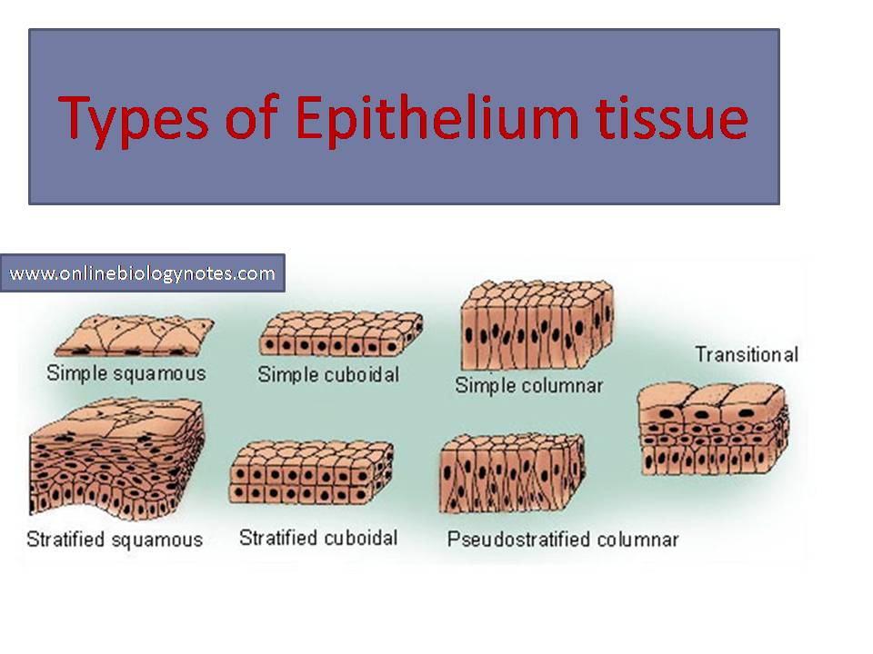
Types of epithelial tissue: simple, compound and specialized ...

The proximal airways represent a pseudostratified columnar ...
Columnar Stock Illustrations – 392 Columnar Stock ...

Lab 2 Epithelial tissue | Histology
Pseudostratified columnar epithelium

What is Epithelial Tissue Different Types of Structure ...
Pseudostratified Columnar Epithelium | Histology, Anatomy & Types

Illustration of Epithelium Types - Stock Image - F031/5310 ...

Pseudostratified Columnar Epithelium
Pseudostratified columnar epithelium Definition, Structure ...

Chapter 2, Page 2 - HistologyOLM
Pseudostratified Columnar Epithelium - AnatomyZone
Comments
Post a Comment