41 holter monitor 5 lead placement diagram
5. Please allow for approximately 30 minutes of testing time. A Holter monitor records the electrical activity of your heart (EKG) for a continuous period of time, usually 24 hours. This test can assist your physician in determining if the symptoms you are feeling are being caused by your heart rhythm. Jan 8, 2020 — Hi, This is another article to guide the users for ECG lead electrodes/cables placement · Monitoring Electrocardiogram- Real time display of the ...
Holter monitor placement. STUDY. PLAY #1 White lead. 1st intercostal space mid clavicular on left ... #4 Brown lead. 4th intercostal space right sternal border #5 Red lead. Lower left at the last rib just anterior of the mid axilary. YOU MIGHT ALSO LIKE... 14 terms. Holter Monitoring ... Diagrams. Flashcards. Mobile. Help. Sign up. Help Center ...
Holter monitor 5 lead placement diagram
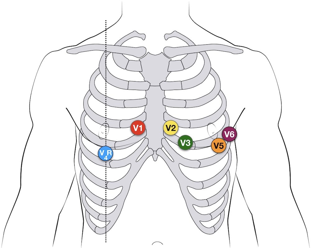
Jun 22, 2014 — www.Empowern.com Tele Lead PlacementHi Guys!This is a quick fun video to show you a little trick about how to place cardiac monitor leads ... Vision 5L Holter Recorder 13 Electrode Placement 3 Channel (5 lead) Electrode Placement Five color-coded leadwires are utilized to create a 3 channel ECG recording. 5 Lead Electrode Placement # Channel Color Placement 1 3- White Below right clavicle, just lateral to the midclavicular line 2 1-, 2- Red Top of the sternum Right sided 12 lead ECG lead placement The most useful lead is V 4 R, which is obtained by placing the V4 electrode in the 5th right intercostal space in the mid-clavicular line. ST elevation in V4R has a sensitivity of 88%, specificity of 78% and diagnostic accuracy of 83% in the diagnosis of RV MI.
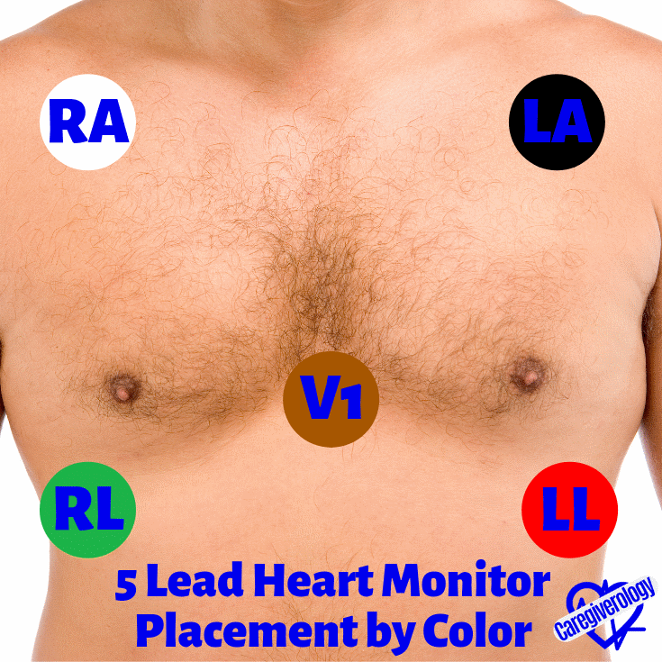
Holter monitor 5 lead placement diagram. Oct 12, 2015 — This video demonstrates how to place electrodes for a 5 lead placement for a cardiac/telemetry monitor for a patient to wear during a ... In the five-lead system, the electrode positions generally remain constant. • Place the right arm (RA) electrode near the right shoulder, close to the junction ... 12 Lead utilizes a 10-wire ECG lead set that can monitor 12 ECG vectors (I, II, III, aVR, aVL, aVF, V1, V2, V3, V4, V5, and V6) simultaneously. The recommended AHA lead placement for a View 12 card is as follows. Holter Monitor 5 Lead Placement Diagram, free sex galleries ekg lead ii placement nursing pinterest, strikani do kalhotek samovolny vyron semena, placement cartoons illustrations
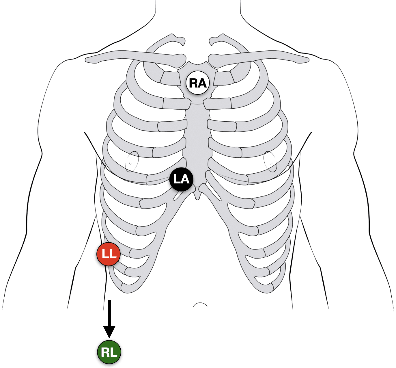
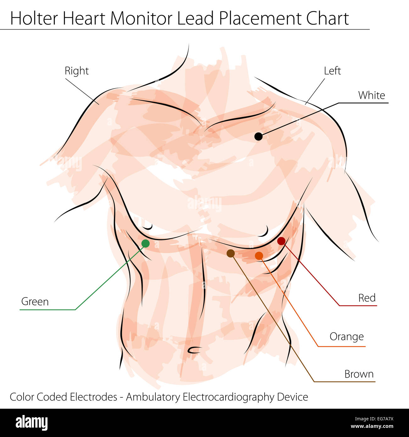
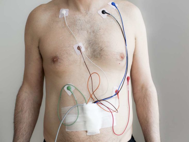
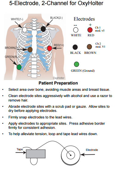
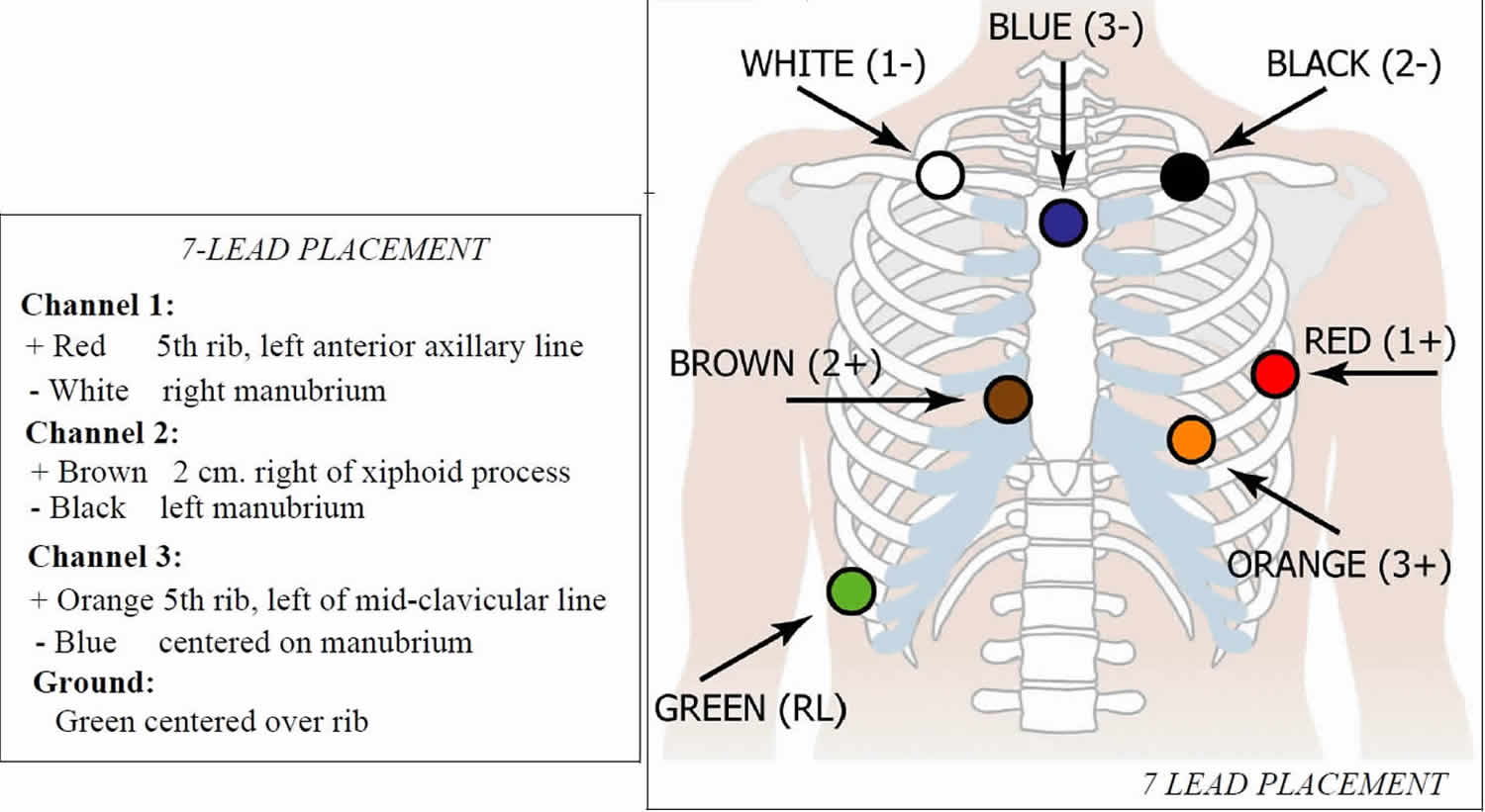
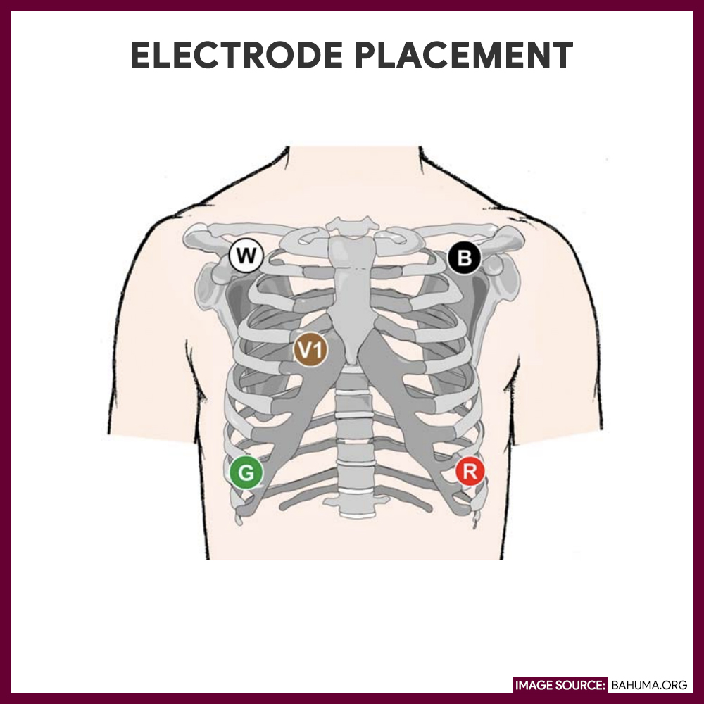
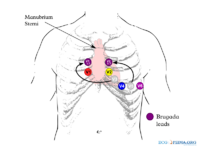
Attached the lead wires to the appropriate electrodes according to the EASI Lead Placement diagram. 5. Place a piece of medical tape over the lead and elec-trode for additional security. This can minimize noise dramatically as the patient moves about their daily ac-tivities. 6. Connect the leads to the Holter monitor and move Although it's referred to as the 12 Lead placement, an ECG only uses ten electrodes for reading. Some electrodes form a pair, which provides this tool with two Leads. The electrodes are self-sticking pads containing a conducting gel at their centers. On the other side, the electrodes snap onto the ECG's or heart monitor's connected cables. 4 lead Holter monitor placement. Appenix 2 - Placement of holter monitor electrode A Holter monitor is a small non-invasive ambulatory, portable ECG machine used to record the heart's. 5 Lead & 12 Lead Placements put somebody onto a cardiac monitor but you cannot remember lead placement, most Image: 5 lead connection box diagram . Philips Holter with Zymed Algorithm Note: Accurate placement and care in proper hookup techniques are absolutely critical for Holter leads. Electrode Placement E (Brown) Level of 5th intercostal space, midsternum A (Black) Same level as E and I, left mid-axillary line S (Red) Top of sternum, manubrium I (White) Same level as E and A, right mid ...
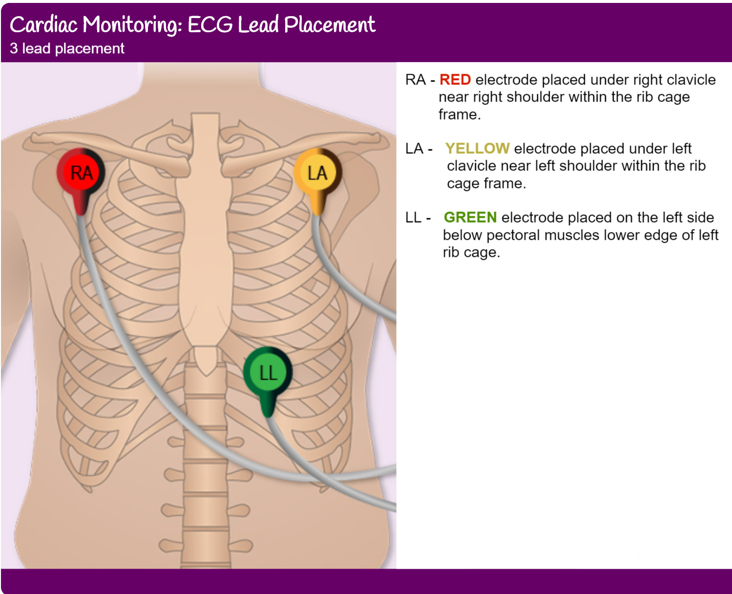
The normal sinus rhythm, as seen in the monitoring to 12 cables. 5-Lead Proper placement of the electrodes is critical. The placement of the electrodes for ECG monitoring is standardized to ensure that the information collected is accurate and can be compared with other occasions recordings. 3 lead ECG cable Placement (there are two ways) Way 1. Monitors one of the three leads: RA: placed the red electrode within the frame of rib cage,right under the clavicle near shoulder( see chart in follow picture) LA: the yellow electrode is placed below left clavicle, which is in the same level of the Red electrode The Holter monitor ambulatory electrocardiography (ambulatory ECG), an ECG recording device. The Holter monitor runs on batteries and can be placed in a pocket or pouch worn around your neck or waist. Holter monitor has 5 to 7 wires called leads. The leads attach to metal disks called electrodes (small conducting patches), which are stuck onto ... Connect cable into monitor, the slot on the cable end only allows cable to be inserted one way. Monitor will beep when cable is properly connected. (The monitor will not turn on unless cable is plugged in.) 5. To turn on the Holter push any one of the keypad buttons.
ECG Lead positioning • LITFL • ECG Library Basics
The Holter monitor continuously monitors and records all heartbeats, detecting irregular rhythms over ... Refer to diagram for proper lead placement The white, green, red and black leads should be proportionally distant from the brown lead and each other. ... 5 • Attach connector portion of the lead wires to the monitor. • Slide the battery ...

ECG - Knowledge @ AMBOSS
The Braemar DL900 3 Channel 5L Holter Cable, 26" - item #350-0290-03, item #CA350029003 is fashioned from the whighest quality medical-professional-grade materials and is designed for use with the DL900 Series Cardiac Event Monitor (sold separately). This 3 channel, 5 lead holter cable also fits the Burdick Mortara 4250 Recorder (sold ...

Diagram For You
Holter Hi-Res / Vector Resting Stress E RL LL RA LA I V4 6 M H Two Channel, 5 Electrode B C A E D B C E A F D G E B C A E D F G B C A D F G RA RL LL LA V1 V2 V3 V4 V5 V6 A3 A1 A2 A1 A2 V6RV5R V4R RA V3R RL LL LA V4 V1 V2 V3 V5 V6 V1 V 5 V6 RL LL RA LA 4 V2 V3 Modi ed Mason-Likar Electrode Placement AHA Label IEC Label Electrode Location V1 (red ...

The Ultimate 12-Lead ECG Placement Guide (With Illustrations)
Start studying Holter 7-lead placement. Learn vocabulary, terms, and more with flashcards, games, and other study tools.

Electrocardiogram 1: purpose, physiology and practicalities ...
The Holter monitor is a type of portable electrocardiogram (ECG). It records the electrical activity of the heart continuously over 24 hours or longer while ...

Holter Monitor Explained by a Cardiologist • MyHeart
Jan 16, 2019 · Appenix 2 - Placement of holter monitor electrode A Holter monitor is a small non-invasive ambulatory, portable ECG machine used to record the heart's. 5 Lead & 12 Lead Placements put somebody onto a cardiac monitor but you cannot remember lead placement, most Image: 5 lead connection box diagram . Guidelines for Holter Monitor Hook-Up.
An image of a holter heart monitor lead placement chart Stock ...
Appenix 2 - Placement of holter monitor electrode A Holter monitor is a small non-invasive ambulatory, portable ECG machine used to record the heart's. 5 Lead & 12 Lead Placements put somebody onto a cardiac monitor but you cannot remember lead placement, most Image: 5 lead connection box diagram.
DR181 Digital Holter Recorder Operator's Manual
5-lead ECG Placement. Ok, let's say you are on your cardiac unit and you're admitting a patient who now needs continuous cardiac monitoring. Other names for this include placing a patient "on the monitor," "monitored," or on telemetry. Knowing 5 lead ECG placement is critical to a fast and efficient admission.
24-hour Holter monitoring: Uses, results, and what to expect
5 Lead & 12 Lead Placements ... to put somebody onto a cardiac monitor but you cannot remember lead placement, ... Image: 5 lead connection box diagram.
TIPS FOR OPTIMIZING ECG RELATED MONITORING ECG electrodes are ...
I have never seen Holter leads placed as described below, but you should check with your Holter manufacturer guidelines. According to ACC guidelines: 2 channel monitor (5 lead wires)
![â–· 12 Lead Placement guide with diagram [VIDEO]](https://aimcardio.com/wp-content/uploads/2020/08/12-leads-resting-ECG-electrode-placemnet.jpg)
â–· 12 Lead Placement guide with diagram [VIDEO]
Apr 20, 2020 · 5-lead monitoring is the same as 3-lead monitoring, but with two additional electrodes that enable the monitoring of extra leads and help improve ST elevation readings (Cables and Sensors 2016). It is able to monitor the leads I, II, III, aVR, aVL, aVF and V (Phillips 2008). The normal sinus rhythmm, as seen in 12-lead monitoring. 5-Lead Placement

Sensor validation protocol at the left side and sensor ...
Right sided 12 lead ECG lead placement The most useful lead is V 4 R, which is obtained by placing the V4 electrode in the 5th right intercostal space in the mid-clavicular line. ST elevation in V4R has a sensitivity of 88%, specificity of 78% and diagnostic accuracy of 83% in the diagnosis of RV MI.
DR181 Digital Holter Recorder Operator's Manual
Vision 5L Holter Recorder 13 Electrode Placement 3 Channel (5 lead) Electrode Placement Five color-coded leadwires are utilized to create a 3 channel ECG recording. 5 Lead Electrode Placement # Channel Color Placement 1 3- White Below right clavicle, just lateral to the midclavicular line 2 1-, 2- Red Top of the sternum
ER920W Ambulatory Heart Monitor User Manual _Rev09 Braemar .
Jun 22, 2014 — www.Empowern.com Tele Lead PlacementHi Guys!This is a quick fun video to show you a little trick about how to place cardiac monitor leads ...

5 Lead & 12 Lead Placements - ECG Lead Placement - Normal ...
Never Underestimate the Importance of a Great Hookup ...
FUSION Ambulatory Arrhythmia Monitoring Device User Manual ...
Heart Monitor Basics and Troubleshooting - Caregiverology
3 Leads ECG Cable and Placement | YQF Medical Cable
Holter Monitor
Holter monitor uses, instructions, preparations and Holter ...

Holter Heart Monitor Lead Placement Chart Stock Vector ...

Understanding Technology and Equipment | Thoracic Key

ECG Lead Placement Guide for 12 Lead ECG | Numed Healthcare
Right Arm Left Arm
2017 ISHNE-HRS expert consensus statement on ambulatory ECG ...

Lead Placement Stock Illustrations – 16 Lead Placement Stock ...

Holter monitors

Practice Standards for Electrocardiographic Monitoring in ...
ECG Lead positioning • LITFL • ECG Library Basics

Practice Standards for Electrocardiographic Monitoring in ...

EKG Basics – Brown Pediatrics

12-Lead ECG Placement | Medical assistant classes, Emergency ...

3 Leads 12 Channels System 24 Hours Dynamic Ecg Holter Monitor Ecg - Buy Holter,Ecg Holter,Holter Monitor Ecg Product on Alibaba.com
Holter Monitoring - Nursing Responsibilities and Care Plan ...

Lead ECG Cable/Electrode Three(3),Five(5),Ten(10)
10002 RhythmStar User Manual RhythmStar Operator's Manual.pub ...

PC Based Resting ECG & EKG Systems Nasiff CardioCard ...
Operator Manual
Basics - ECGpedia
ECG Lead Placement Proc 7664revA.pptx
Comments
Post a Comment