41 drag the labels onto the diagram of the cns meninges.
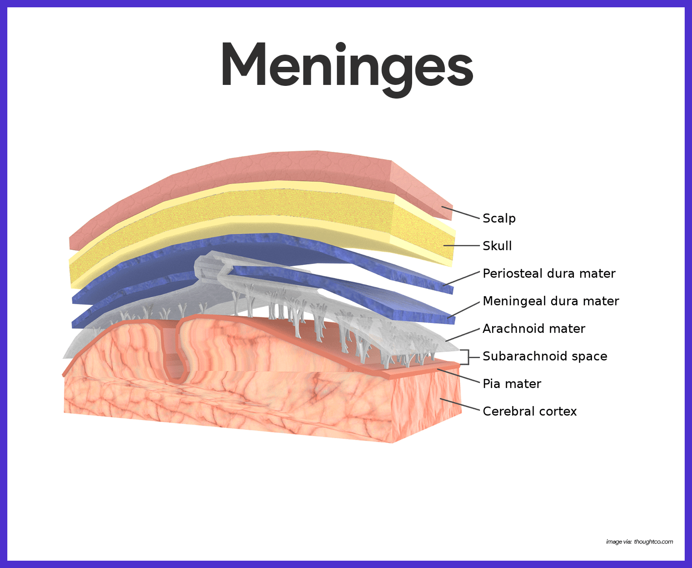
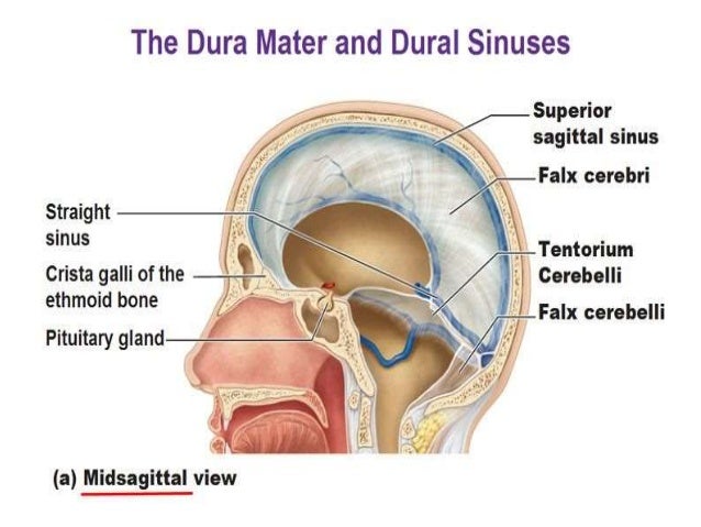
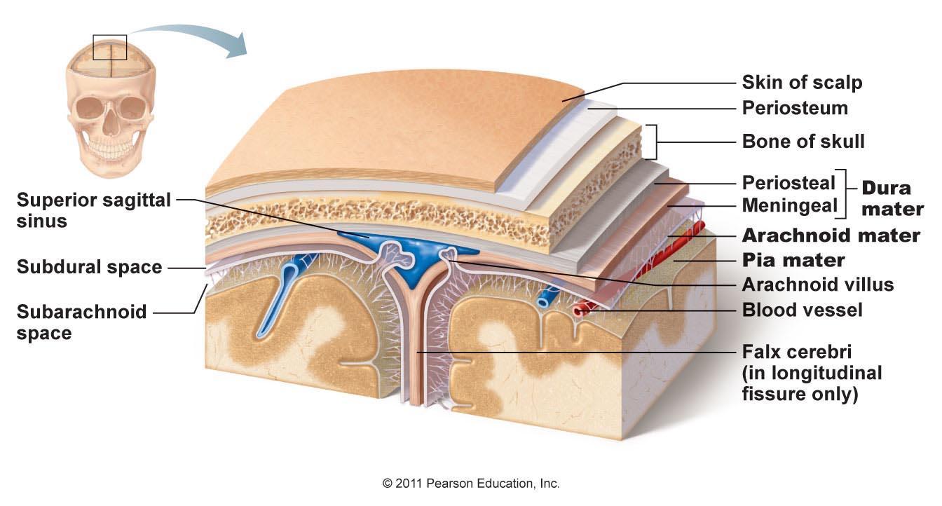
In anatomy, the meninges are the three membranes that envelop the brain and spinal cord. In mammals, the meninges are the dura mater, the arachnoid mater, and the pia mater. Cerebrospinal fluid is located in the subarachnoid space between the arachnoid mater and the pia mater. The meninges are the membranes that surround and protect the brain and the spinal cord. In mammals, the meninges have three layers: the dura mater, the arachnoid mater, and the pia mater. In the space between the arachnoid mater and the pia mater (called the "subarachnoid space"...
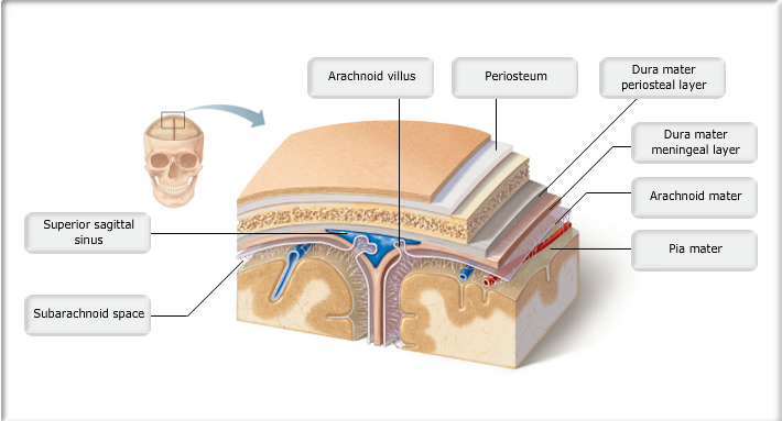
Anatomy and Physiology. Anatomy and Physiology questions and answers. Part A Drag the labels onto the diagram to identify the cranial meninges and associated structures. Reset Hel Subdural space Pia mater Subarachnoid space Dural sinus Cranium Dura mater (meningeal layer) Dura mater (periosteal layer) Arachnoid mater Cerebral cortex QUID Submit Request Answer.
Drag the labels onto the diagram of the cns meninges.
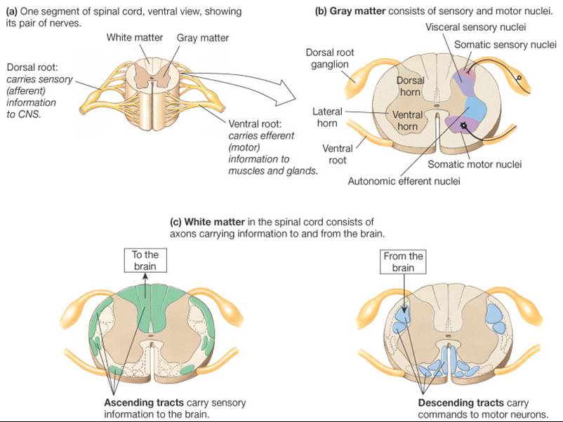
Jul 01, 2019 · The meninges functions primarily to protect and support the central nervous system (CNS). It connects the brain and spinal cord to the skull and spinal canal. The meninges forms a protective barrier that safeguards the sensitive organs of the CNS against trauma. It also contains an ample supply of blood vessels that deliver blood to CNS tissue. We review their content and use your feedback to keep the quality high. 100% (11 ratings) Answer The label is indicated from RIGHT SIDE of image to …. View the full answer. Transcribed image text: Part A Drag the labels onto the diagram to identify the spinal nerve roots and meninges Reset Help Ventral Pia mater Meninges Dorsal root Dura mater. The meninges are the fibrous covering of the central nervous system (CNS) which Meningeal Cell Types. The meninges contain two compartments: the leptomeninges (collective term for pia and (D) Immunofluorescence image of human fetal leptomeninges in the Sylvian sulcus labeled with CRABP2...
Drag the labels onto the diagram of the cns meninges.. Drag the labels onto the diagram to identify the cranial meninges and associated structures. 1. dura mater 2. subarachnoid space 3. pia mater 4. cerebral cortex 5. cranium 6. periosteal cranial dura 7. dural sinus 8. meningeal cranial dura 9. subdural space 10. arachnoid mater. The central nervous system (CNS) consists of the brain and the spinal cord. Our brains have two primary functions, which are to control behavior and to regulate the body's physiological processes. However, the brain cannot do this alone as it needs to receive information from the body's sense... The central nervous system (CNS) is the part of the nervous system consisting of the brain and spinal cord. As such, the olfactory epithelium is the only central nervous tissue in direct contact with the environment, which opens up for therapeutic treatments. Label the meninges of the brain and spinal cord.Part ADrag the labels onto the diagram of the CNS meninges.Central canalAqueduct of SylviusArachnoid villusCranial vein valves. 4/11/2017Chapter 910/10CorrectScore Summary:Your score on this assignment is 97.1%.You received...
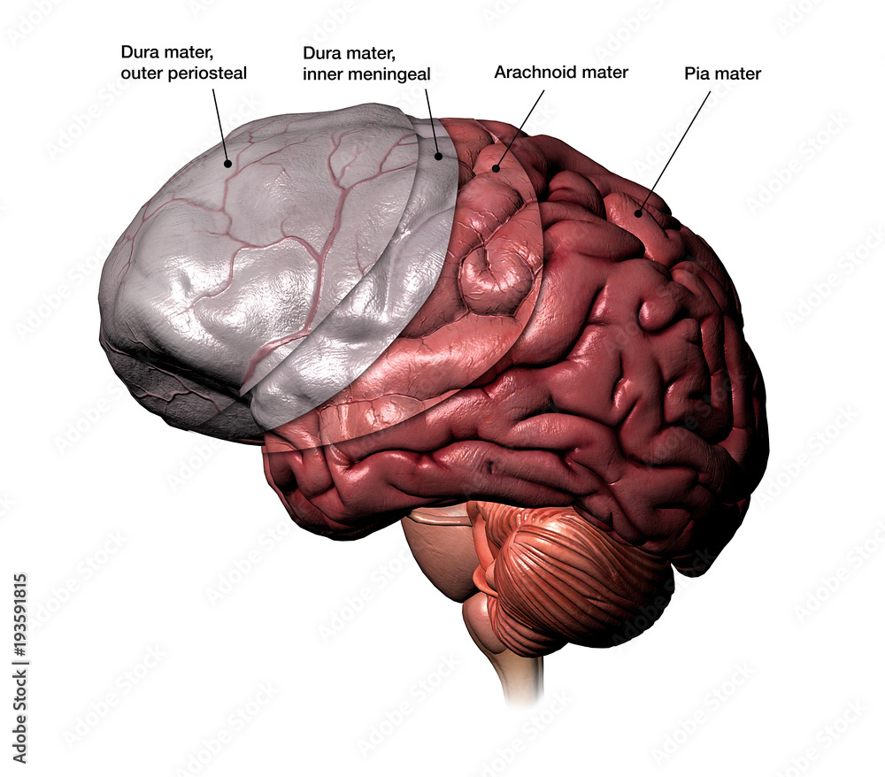
drag-the-labels-onto-the-diagram-to-identify-the-classes-of-epithelia. Uploaded on Aug 09, 2021. Download Presentation. pia mater onto the brain ct covering arachnoid mater tough ct double layer outer periosteal, inner meningeal connective tissue coverings of. External cover that protects brain & spinal cord Ń Extensions into cranial cavity limit movement of the brain `. Meninges that surround the CNS consist of an outer fibrous sheet of dura mater (pachymeninx) that is also the inner periosteum of the skull. As the foramina of Magendi and Luschka develop, one continuous CSF system evolves. Due to the lack of arachnoid granulations during foetal life, it is most... Drag and drop the text labels onto the boxes next to the heart diagram. 1 pulmonary ventilation the total exchange of air usually measured in litersminute and 2 alveolar ventilation the effective ventilation of the alveoli in which gas exchange with the blood actually takes place.
The meninges refer to the membranous coverings of the brain and spinal cord, comprised of the dura, arachnoid and pia mater. Provide a supportive framework for the cerebral and cranial vasculature. Acting with cerebrospinal fluid to protect the CNS from mechanical damage. 2- Histology of spinal cord-CNS-Second year-New edition 2021. Neurology | Anatomy & Function of the Cerebellum. Cns central nervous system 7. Can you name the label the parts of the neuromuscular junction. Drag and drop the descriptive labels of events into the correct sequence at the chemical synapse. Drag and drop the descriptive labels of events into the correct sequence at the chemical synapse. 1 4 The Somatic Nervous System Neuroscience Canadian 1st Edition. Label the parts of the neuromuscular junction. Cns central nervous system 7. Drag the labels onto the diagram to...
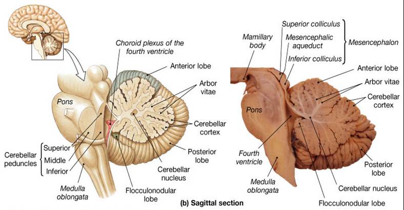
Lab Week 3: Spinal Cord and Brainstem – Rehab 551 Lab
Meninges. The brain and the spinal cord Spinal cord The spinal cord is the major conduction pathway connecting the brain to the body; it is part of the CNS. of the meninges, and the etiology can be elicited by examining CSF, which is contained within the subarachnoid space.
Anatomy of the Head and Neck | SpringerLink
Reset Help central cand matrix Group 2 lacuna Group 2 Group 2 osteocyte in lacuna Group 2 C chondrocyto Group 2 bono (osseous tissue) Group 1 Bone tissue differentiates from other tissues because of the mineralized extracellular matrix. This tissue provides support and protection due to the...

Viral Meningitis - Brain, Spinal Cord, and Nerve Disorders ...
In November, millions of Monarchs fall like bright, golden rain onto the forests in the mountains of central Mexico. Their long journey to Mexico is thought to be one of the most amazing events in the whole of the American continent.
Neuroanatomy Online: An Open Access Electronic Laboratory for ...
After each piece of the lagging stand is complete it is released from dna polymerase. Na is entering the cell. 3 3 Eukaryot...

Solved Part A Drag the labels onto the diagram to identify ...
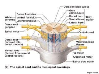
Spinal nerves Part of the peripheral nervous system 31 pairs attach through dorsal and ventral nerve roots Lie in intervertebral foramina. Protection: Bone Meninges CSF (cerebrospinal fluid) 3 meninges: dura mater (outer) arachnoid mater (middle) pia mater (inner) 3 potential spaces epidural...

A&P 2 Lab 5 HW Flashcards | Quizlet
The Central Nervous System (CNS) consists of: The spinal cord Integrates and processes information Can function with the brain Can function independently. Chapter 14 - The Nervous System: The Spinal Cord and Spinal Nerves $100 $200 $300 $400 $500 $100$100$100 $200 $300...
Anatomy and Physiology Lab I†on OpenALG
Meninges-brain interface: signals from the meninges regulate development of the CNS 1041. Infiltration of the meninges is such a common complication of acute leukaemia that routine treatment of acute leukaemia in childhood includes regular prophylactic measures to prevent its occurrence.

294 Meninges Stock Photos and Images - 123RF
The central nervous system (CNS) consists of the brain, spinal cord, spinal fluid and neurons, which transmit signals from the brain to other body Efferent neurons are mainly located in the peripheral nervous system, but their cell bodies orginate in the CNS. Many incoming signals from the CNS...
Nervous System Anatomy and Physiology - Nurseslabs
Drag the labels onto the diagram to identify the spinal nerve roots and meninges. look at pic Drag the labels onto the diagram to identify the parts of the spinal cord (transverse section, showing white matter).

dura mater, the arachnoid mater, and the pia mater | Dura ...
The meninges are the fibrous covering of the central nervous system (CNS) which contain vastly heterogeneous cell types within its three layers (dura, arachnoid, and pia). These results identify a private source for meningeal B cells. which may help maintain immune privilege within the CNS.

9 BRAIN ideas | brain, dura mater, brain anatomy
Drag the labels onto the diagram to identify the cranial meninges and associated structures. Drag the labels to identify the landmarks and features on one of the cerebral hemispheres. Drag the labels onto the diagram to identify the origins of the cranial nerves (I - VI).

Îωτιαίος μυελός - νωτιαία νεÏÏα. spinal cord | Spinal cord ...
The central nervous system is the supreme command center of the human body. Learn about its anatomy and function now at Kenhub! The CNS consists of two organs which are continuous with each other; the brain and spinal cord. They are enveloped and protected by three layers of meninges...

Drag the appropriate labels to their respective targets ...
Feb 05, 2019 · View Screen Shot 2019-02-05 at 8.26.00 PM.png from BSC 2086L at University of South Florida. Drag the labels onto the diagram to identify the cranial meninges and associated structures.
Meninges, bbb, csf, icp, brain edema, hydrocephalus
The central nervous system (CNS) consists of the brain and spinal cord. This body system is responsible for integrating and coordinating the activities of the entire body. Overview. The central nervous system can be thought of as the coordination and integration system within organisms.
Associate Degree Nursing Physiology Review
Drag the arrows onto the diagram below to indicate the direction that DNA polymerase III moves along the parental (template) DNA strands at each of the two replication forks. 5 hours ago Art-labeling Activity: Brain, Cranium, and Meninges (Close-up View of Cranial Meninges) Part A Drag the labels...
Anatomy and Physiology 1 Chapter 12 Flashcards - Easy Notecards
The meninges are the fibrous covering of the central nervous system (CNS) which Meningeal Cell Types. The meninges contain two compartments: the leptomeninges (collective term for pia and (D) Immunofluorescence image of human fetal leptomeninges in the Sylvian sulcus labeled with CRABP2...

lab 7 (exercise 14) Flashcards | Quizlet
We review their content and use your feedback to keep the quality high. 100% (11 ratings) Answer The label is indicated from RIGHT SIDE of image to …. View the full answer. Transcribed image text: Part A Drag the labels onto the diagram to identify the spinal nerve roots and meninges Reset Help Ventral Pia mater Meninges Dorsal root Dura mater.

lab 7 (exercise 14) Flashcards | Quizlet
Jul 01, 2019 · The meninges functions primarily to protect and support the central nervous system (CNS). It connects the brain and spinal cord to the skull and spinal canal. The meninges forms a protective barrier that safeguards the sensitive organs of the CNS against trauma. It also contains an ample supply of blood vessels that deliver blood to CNS tissue.

Nervous System Anatomy and Physiology - Nurseslabs

A&P2 Lab 3 HW Flashcards | Quizlet
Associate Degree Nursing Physiology Review

Meninges & CSF - Histology | Draw it to Know it
From outermost to innermost, what are the names and the ...
Spinal cord: Ascending and descending tracts | Kenhub
SAY: Welcome to Module 1: Anatomy & Physiology of the Brain ...

Case Study - Stroke A 77-year-old woman was cooking in the ...

A&P2 Lab 3 HW Flashcards | Quizlet
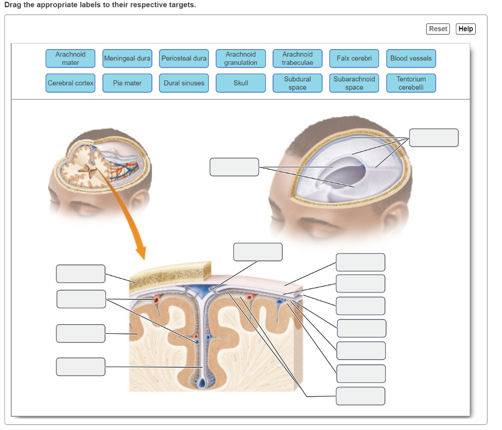
Solved Drag the appropriate labels to their respective ...
Anatomy and Physiology Lab I†on OpenALG

JaypeeDigital | eBook Reader

Brain
Associate Degree Nursing Physiology Review
Human Brain Revealing Meninges Layers with Labels Stock ...
Spinal cord: Ascending and descending tracts | Kenhub
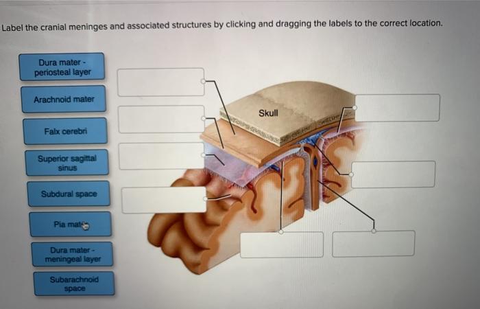
Solved Label the cranial meninges and associated structures ...
Spinal cord and spinal nerves lab

A&P2 Lab 2 HW Flashcards | Quizlet

12.2 Nervous Tissue – Anatomy & Physiology

Pacific Medical Training - Nervous System
The Nervous System
The Brain, Cranial Nerves, and Sensory and Motor Pathways
Comments
Post a Comment