40 ap psychology eye diagram
Eye Structure Psychology. Here are a number of highest rated Eye Structure Psychology pictures upon internet. We identified it from trustworthy source. Its submitted by government in the best field. We acknowledge this kind of Eye Structure Psychology graphic could possibly be the most trending topic later we ration it in google benefit or ... AP Psych Eye Diagram STUDY PLAY Cornea Clear, curved bulge of tissue on the front of the eye that bends light rays to begin focusing them and protects the eye (lots of nerve endings) Iris Ring of muscle tissue that forms the colored portion of the eye; surrounds the pupil and controls its size Pupil
AP Psychology 🧠 Bookmarked 10.9k • 322 resources See Units Vocabulary neuron myelin sheath threshold neurotransmitters agonist dendrites action potential all-or-none response reuptake antagonist axon refractory period synapse endorphins Neural Firing Parts of the Neuron Neurons are our body's nerve cells which make up the nervous system.

Ap psychology eye diagram
1- Complete the Unit 1 Progress Check on AP Classroom 2- Complete the Unit 2 Progress Check on AP Classroom 3- Complete three viewing guides for review videos on YouTube by College Board (see TEAMs for directions and viewing guide) Virtual Learning Week #2 Assignments Monday, March 30 - Friday, April 4 Step Two: The Light Channeled within the Eye Once the light hits the eye it goes through a variety of structures. Take a look at the diagram below. The white part of our eye is called the cornea and basically protects and helps reflect light. The light goes through a hole in our eye called a pupil . ear diagram.pdf - 1. Ear Canal 7. Cochlea 2. Ear Drum 8.... School Parkway North High School Course Title AP PSYCHOLOGY 15575 Uploaded By JusticeSummer1306 Pages 2 This preview shows page 1 - 2 out of 2 pages. View full document 1. Ear Canal 2. Ear Drum 3. Hammer 4. Anvil 5. Stirrup 6. Semicircular Canals 7. Cochlea 8. Auditory Nerve 9.
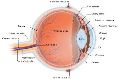
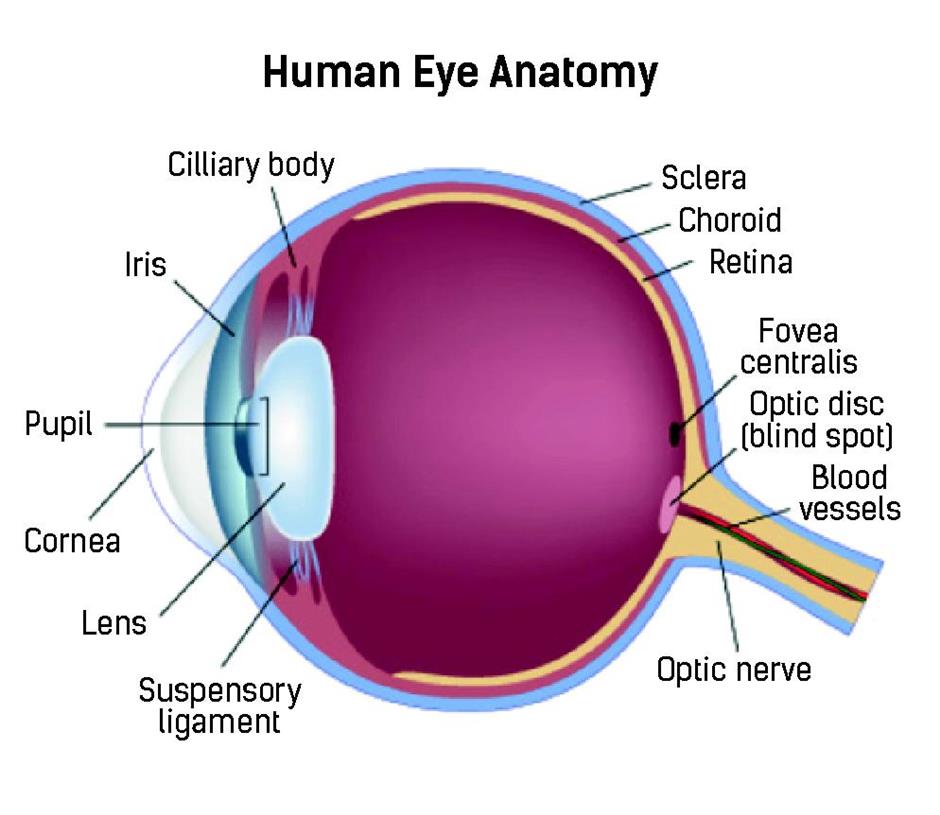
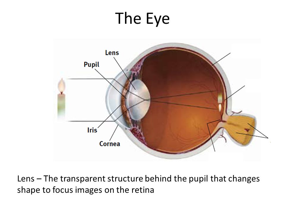
Ap psychology eye diagram. (illustrations, diagrams, clipart) are extensive, varied & appropriate to the task and content Evidence of personal meaning making is clear on the left side Analysis is strong (captions, labels, summaries are included) The student gave care to ensure neatness and legibility Student-initiated extras are included Details -Quiz Diagram on Eye & Ear -Review thus far HW= Module 21 (Due Wednesday 23) wednesday 10/23 -Rotation Activity with Other Senses - Touch, Pain, Taste, Smell HW = Makae sure all your reading and notes are done at this point thursday 10/24 - parent teacher CONFERENCES Conference Sign-Ups Review DN: How are you going to study for this test? of light entering the eye. Lens: The lens is a clear part of the eye behind the iris that helps to focus light, or an image, on the retina. Macula: The macula is the small, sensitive area of the retina that gives central vision. It is located in the center of the retina. Optic nerve: The optic nerve is the largest sensory nerve of the eye. Label and shade the diagram of the eye below (only label and shade the bolded parts). Describe the functions of each eye part in the chart that follows. Eye ...4 pages
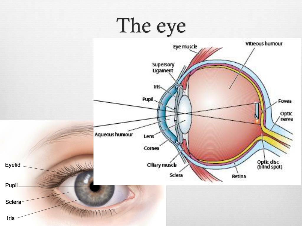
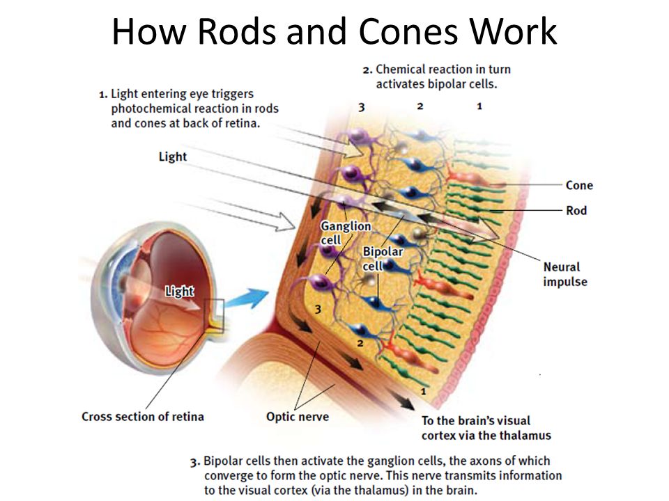
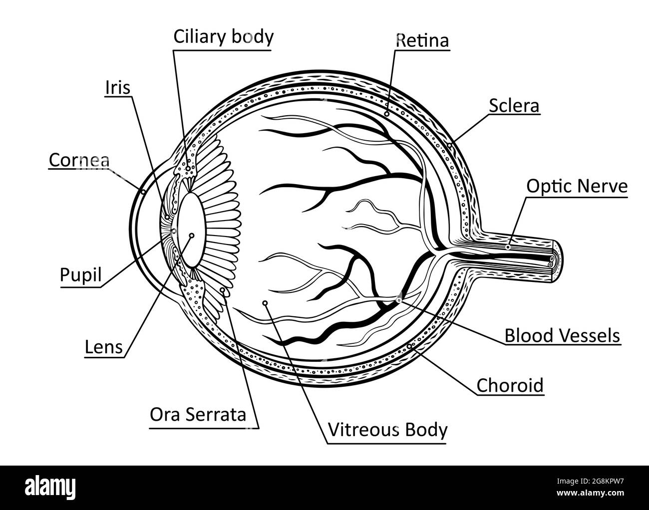
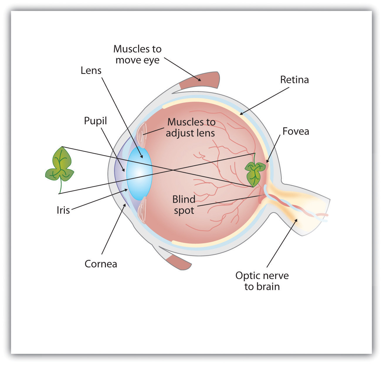
AP Psychology - Unit 4 Assignment ... Draw a diagram of the eye. Label and explain the function of the iris, lens, pupil, cornea, retina, fovea, blind spot, and optic nerve. 5. What is transduction and how does this process occur in the photoreceptors of the eye and cochlea of the ear? 6. Draw a diagram of the ear. along the back of the eye and it contains the rods, cones, bipolar and ganglion cells. Use "red tin" as your mnemonic and imagine that the back of your eye is covered with red tin. Fovea: is a spot in the eye that is directly behind the lens. There is a very high concentration of cones in this area which means that images t Eye Diagram Retina to Brain Diagram F. Describe the vision process, including the specific nature of energy transduction, relevant anatomical structures, and specialized pathways in the brain for each of the senses. G. Explain common sensory conditions. This Is Not Yellow Do Those Glasses Really Fix Colorblindness? 3.4 Visual Perception Module 9 Flip It Video - Action Potential. Module 10 Flip It Video - The Reflex Arc. Module 10 Flip It Video - Structure of the Nervous System. Module 11 Flip It Video - Limbic System. Module 12 Flip It Video - Structure and Function of the Cortex. Module 13 Flip It Video - Split-Brain Research.
Eye Diagram Handout Author: National Eye Health Education Program of the National Eye Institute, National Institutes of Health Subject: Handout illustrating parts of the eye Keywords: parts of the eye, eye diagram, vitreous gel, iris, cornea, pupil, lens, optic nerve, macula, retina Created Date: 12/16/2011 12:39:09 PM A 360 Moving Diagram of the Eye Parts (choose what type of section you would like to see and then press the play button underneath the image) Find Your Blind Spot Activity Interactive Measure of... AP Psychology Study Guide ... take to ring a bell) nerve leaves the eye oMedulla - vital organs (HR, BP) o Pons - sleep/arousal (Ponzzzzzz) • Midbrain o Reticular formation: attention (if you can't pay attention, You R F'd) • Forebrain: higher thought processes Best AP Psychology Review Book for Low-Scoring Students: Cracking the AP Psychology Exam, 2020 Edition. Cost: $18 for print, $13 for digital Written by The Princeton Review, this is by far the best book for learning test-taking strategies for the AP Psychology test.The content is high quality as well, but it's not as easy to study from if you don't have much time on your hands.
20 Questions Tuesday: 419 - Vision — 20 Questions Tuesday
AP Psychology In addition to the information in this study guide, you are also responsible for all of the content in textbook (Modules 16-21), all information from class notes/discussions, all handouts and graphic organizers. It's AP - it's all fair game Terms & Concepts All Key Terms & Concepts to Remember on page 214 (Modules 16-21)
AP Psychology Ch. 3 Flashcards | Memorang
38. $2.00. Zip. This pack features high-quality anatomical diagrams of the human eye and ear and is ideal for middle school life science or high school biology students. Included are two, one-page worksheets and answer keys. The eye diagram worksheet asks students to first match identified parts of the eye with t.

AP Psych Eye Diagram Diagram | Quizlet
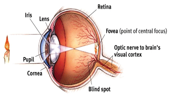
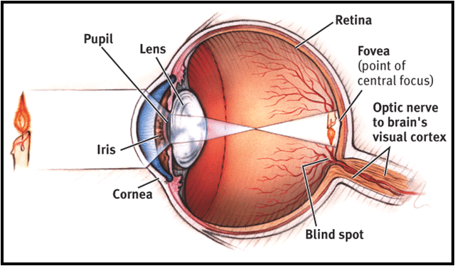
Image Courtesy of Myers AP Psychology Textbook - 2nd Edition 1. First, light passes through the cornea, a thin tissue that protects the eye and bends light to provide focus. 2. Next, light passes through the pupil, a small opening controlled by the iris.

Pharmaceutics | Free Full-Text | Viral-Vector-Delivered Anti ...
3. Diagram out a close up of the Photoreceptors with explanation on how these w. o. rk. 4. Diagram out the steps in the process of Transduction. 5. Diagram out an example of the Opponent- Process Theory with afterimages. 6. Diagram out the visual pathways of the eye to the optic nerves, to the brain.
AP Psychology on Twitter: "Eye diagram. #APPsych https://t.co ...
AP Psychology Please check back here for more information, assignments, notes, etcetera. Sign up for REMIND by texting the code @acpsych to the number 81010.
PPT - Perception PowerPoint Presentation, free download - ID ...
Label these ear parts. Learning: S.T.E.M. It's all happening. Anatomy is the study of the structure of human, animals and plants. Now Introduce your child to anatomy with these 10 free printable anatomy coloring pages. Label Ear Anatomy Diagram Printout.

AP Psych Eye Diagram Diagram | Quizlet
The Forebrain The forebrain consists of the thalamus, hypothalamus, amygdala, and the hippocampus. The hypothalamus, amygdala, and hippocampus make up what we call the Limbic System of your brain. Thalamus The thalamus is located between the cerebral cortex and the midbrain. It is made up of nuclei that receive different sensory and motor inputs.

AP PSYCH: Sensation and Perception EYE DIAGRAM Diagram | Quizlet
Bozeman Public Schools Social Studies Standards, AP Psychology Page 1 of 18 Updated January 23, 2010 Bozeman Public Schools Social Studies Curriculum Advanced Placement AP Psychology ... H.9.3 Students label a diagram of the parts of the eye, ear, tongue and skin receptors, explaining the role and operation of each part. Students distinguish ...

Human Eye Ball Anatomy & Physiology Diagram
AP Psychology Eye and Ear. STUDY. Learn. Flashcards. Write. Spell. Test. PLAY. Match. Gravity. Created by. Tamatha73 TEACHER. Terms in this set (51) Light enters the eye through the pupil and reaches the _____, which focuses light on the retina. lens.

Myers' PSYCHOLOGY (6th Ed) Chapter 5 Sensation. The spectrum ...
AP Psychology Eye diagram STUDY Flashcards Learn Write Spell Test PLAY Match Gravity Retina Click card to see definition 👆 The light-sensitive inner surface of the eye, containing the receptor rods and cones plus layers of neurons that begin the processing of visual information. Click again to see term 👆 1/8 Previous ← Next → Flip Space
Vision AP Psych Transduction – converting one form of energy ...
4-2-eye_diagram (4).pdf - | Course Hero 4-2-eye_diagram (4).pdf - School Campbell High School Course Title PSYCHOLOGY AP Uploaded By sweetmelissasbnb Pages 1 This preview shows page 1 out of 1 page. View full document End of preview. Want to read the entire page? Upload your study docs or become a Course Hero member to access this document

Test: AP Psych Eye Diagram | Quizlet
• Bottom-up processing • Top-down processing • Depth perception o Binocular depth cues Retinal disparity Retinal convergence o Monocular depth cues Overlap Gradient/texture Relative size Linear perspective Aerial perspective • Gestalt principles o Closure, proximity, figure-ground, continuity, similarity *see diagram

AP Psych - Sensory Mechanisms: Eyes & Ears
ear diagram.pdf - 1. Ear Canal 7. Cochlea 2. Ear Drum 8.... School Parkway North High School Course Title AP PSYCHOLOGY 15575 Uploaded By JusticeSummer1306 Pages 2 This preview shows page 1 - 2 out of 2 pages. View full document 1. Ear Canal 2. Ear Drum 3. Hammer 4. Anvil 5. Stirrup 6. Semicircular Canals 7. Cochlea 8. Auditory Nerve 9.

Eye and Ear - *w W LCSson l l Truths-9r Rtnuurcc i Diagram-a ...
Step Two: The Light Channeled within the Eye Once the light hits the eye it goes through a variety of structures. Take a look at the diagram below. The white part of our eye is called the cornea and basically protects and helps reflect light. The light goes through a hole in our eye called a pupil .
Phacoemulsification the gold standard of cataract surgery ...
1- Complete the Unit 1 Progress Check on AP Classroom 2- Complete the Unit 2 Progress Check on AP Classroom 3- Complete three viewing guides for review videos on YouTube by College Board (see TEAMs for directions and viewing guide) Virtual Learning Week #2 Assignments Monday, March 30 - Friday, April 4
Chapter 6 Sensation and Perception Flashcards | Chegg.com
Ora Serrata High Resolution Stock Photography and Images - Alamy
Vision AP Psych Transduction – converting one form of energy ...
Structure and Function of the Eyes - Eye Disorders - Merck ...

AP Psychology 2014-2015: The Sensory Processes

The Eye - Science Quiz

Eye diagram: AP Psychology Diagram | Quizlet
Antioxidants | Free Full-Text | Antioxidant Defenses in the ...
Chapter 9: The Human Eye
Net Vision Search Site Map Contact Net Vision Optic Nerve ...
Human Eye Iris Diagram Mammalian Eye - Parts Of The Eye ...

OpTranslate - Home | Facebook

Eye + ear

Retina: Definition & Function - Video & Lesson Transcript ...

AP Psychology Review on Twitter: "Anatomy and Function of the ...

State of the Art: Progress Toward Restoring Vision - Brain ...

Psych Sensation and Perception Myers 20-25 - Quizizz
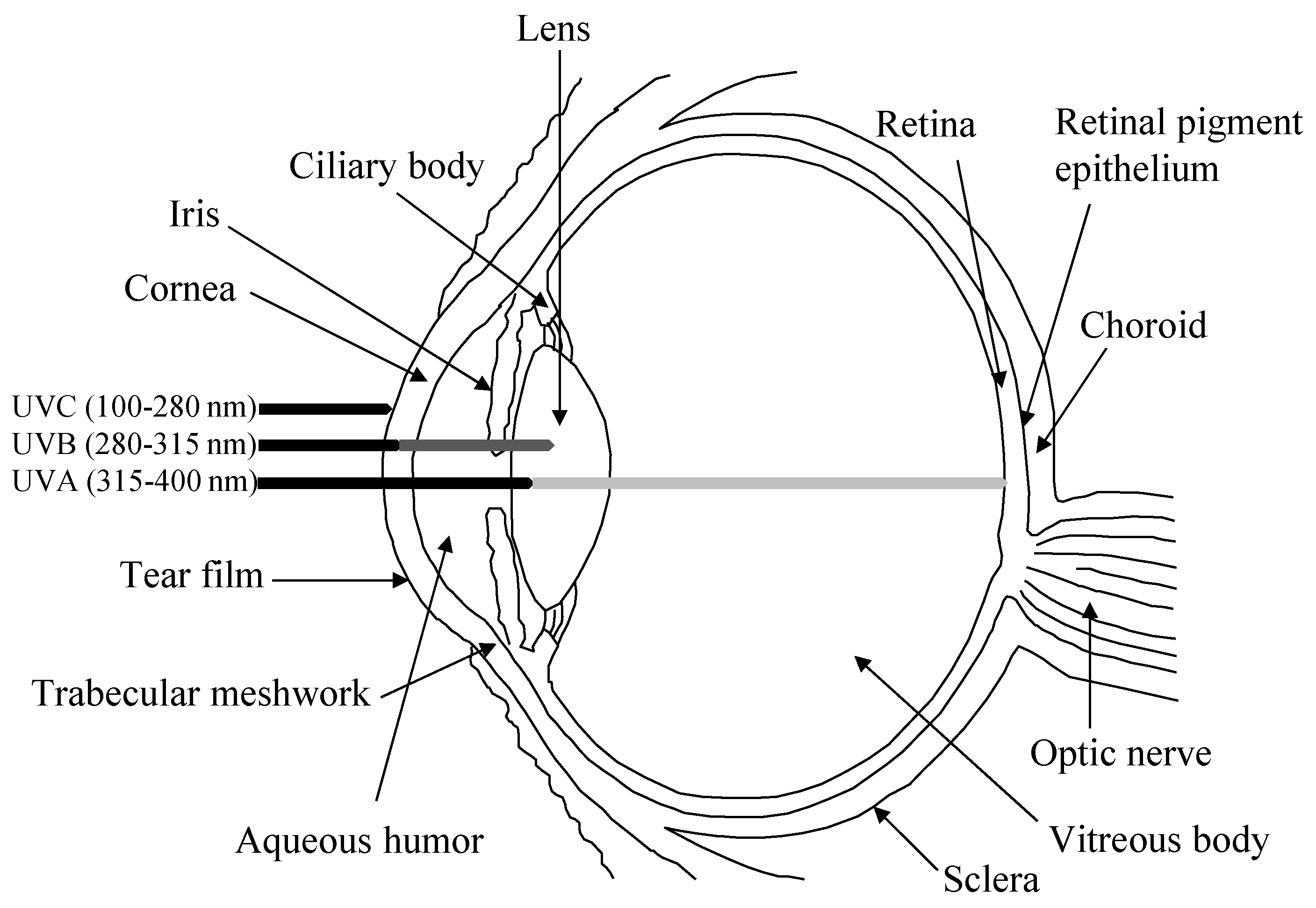
Medical laser safety
5.2 Seeing – Introduction to Psychology – 1st Canadian Edition

Types of Eye Diseases | Study.com
Ms. Scott's A.P. Psychology Summer Assignment due on the ...
Retinal detachment Information | Mount Sinai - New York

Optical Express - Did you know, patients who have waited more ...
AP Psychology chapter 4: Sensation

Lutein and zeaxanthin are... - Energlo Lutein and Zeaxanthin ...
Comments
Post a Comment