39 dog bone diagram
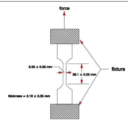
Label Dog Anatomy Diagram Dog Printouts Read the definitions below, then label the dog external anatomy diagram. back - the part of the body between the loin and the withers. brisket - the chest of the dog. carpals - the wrist, the bones of the pastern joint. ASTM dog bone test specimen was designed using Siemens NX 10 CAD software. The test specimens were designed to conform to ASTM D-638, Stan- dard Test Method for Tensile Testing of Plastics, as ...
Atlas of anatomy on x-ray images of the dog. This module of vet-Anatomy is a basic atlas of normal imaging anatomy of the dog on radiographs. 51 sampled x-ray images of healthy dogs performed by Susanne AEB Borofka (PhD - dipl. ECVDI, Utrecht, Netherland) were categorized topographically into seven chapters (head, vertebral column, thoracic limb, pelvic limb, larynx/pharynx, thorax and abdomen ...

Dog bone diagram
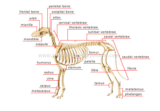
skeleton of a dog. previous. next. caudal vertebrae Bony parts comprising the skeleton of the tail located at the terminal end of the vertebral column. ... Flat skull bone forming the forehead and top of the eye sockets, and articulating especially with the parietal. Speaking of skeletons, a dog has 320 bones in their body (depending on the length of their tail) and around 700 muscles. Muscles attach to bones via tendons. Depending on the breed of dog, they will have different types of muscle fibers. You've probably heard about slow and fast twitch muscle fibers before. Download scientific diagram | ASTM D 638 "dog bone" specimen. from publication: On the Feasibility of Thermoelastic Stress Analysis on Rapid Prototyping Models | This work discusses application of ...
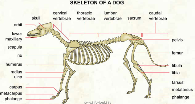
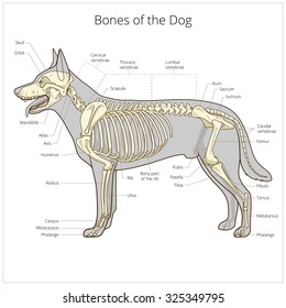
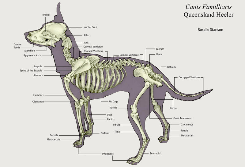
Dog bone diagram. Skeleton of a dog: carnivorous domestic mammal raised to perform various tasks for humans. Skull: bony case of the brain. Cervical vertebrae: bones of the neck. Thoracic vertebrae: the bones forming the dorsal part of the thoracic cage. Lumbar vertebrae: the bones of the lumbar region of the back. Sacrum: the set of sacral vertebrae. Dog anatomy is not very difficult to understand if a labeled diagram is present to provide a graphic illustration of the same. That is exactly what you will find in this DogAppy article. It provides information about a dog's skeletal, reproductive, internal, and external anatomy, along with accompanying labeled diagrams. Dog skeleton. As with any vertebrate animal, the skeleton of a dog has the function of supporting the body for movement and protecting its internal organs. We can divide the canine skeleton into three main sections: Axial skeleton: skull, spine, ribs and sternum bones. Appendicular skeleton: bones of the extremities. The nasal bone is long, slender, and narrow caudally in a dog. The external surface of the nasal bone varies in size and shape. A mucous membrane covers the ventral or internal surface of the dog's nasal bone. For more dog bone-labeled diagrams, you may follow anatomy learner on social media. Mandible of the dog
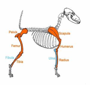
The dog has 321 bones. Regions of a Long Bone Structure of a Long Bone articular cartilage nutrient artery entering nutrient foramen marrow cavity compact bone spongy bone ligament periosteum endosteum physis (epiphyseal plate) physis (epiphyseal plate) metaphysis diaphysis metaphysis epiphysis Hemotopoietic System ~ Bone Marrow and the Blood Cells Includes the bone marrow which is located inside the bones. Three types of blood cells are made in the bone marrow: white blood cells that fight infection, red blood cells that carry oxygen and platelets that are part of the blood clotting process. Special Senses of the Dog ~ Eyes, Ears Dogs have disconnected shoulder bones (lacking the collar bone of the human skeleton) that allow a greater stride length for running and leaping. Bones and Joints of a Dog. The forelegs and hind legs of a dog are as different as human arms and legs: The upper arm on the foreleg is right below the shoulder and is comprised of the humerus bone. This short post will try to cover the dog leg anatomy in detail with labeled diagrams. The leg of a dog consists mainly of the two long bones - tibia and fibula. So, here you will get the detailed anatomy of the leg region of a dog (bones, muscles, and vessels). But, I will also discuss the anatomy of other parts of the dog's hind limb.
Bone Diagram Forehead (Frontal bone) Nose bones (Nasals) Cheek bone (Zygoma) Upper jaw (Maxilla) Lower jaw (Mandible) Breast bone (Sternum) Upper arm bone (Humerus) Lower arm bone (Ulna) Thigh bone (Femur) Collar bone (Clavicle) Toe bones (Phalanges) Ankle bones (Tarsals) Kneecap (Patella) Shin bone The cervical part of dog spine anatomy includes the cervical vertebrae, thick intervertebral discs, part of the spinal cord, and cervical spinal nerves. Now, I will describe the anatomical facts of dog cervical vertebrae with a labeled diagram. In most mammals, you will find seven cervical vertebrae in their vertebral column. 13. the radius and ulna. The dog's radius and ulna run down the front leg to the carpal bones. [adrotate group="7"] 14. The dog's carpal bones. The carpal bones in the front legs form what is the equivalent of the dog's wrist. [adrotate group="7"] 15. The dog's pastern. Electrical Adapters 30 50 Amp Dog Bone Adapter 50 Amp Rv Wiring in 50 Amp Rv Wiring Diagram by admin From the thousands of photos on-line about 50 amp rv wiring diagram, we choices the best libraries along with best quality just for you, and now this images is among pictures selections in this ideal photographs gallery concerning 50 Amp Rv Wiring Diagram.
Dog skeleton with major bone elements labeled (Davis, 1987, p ...
Dogs are digitigrade animals and bear weight on digits II to V, with the main weight bearing occurring on digits III and IV. The sesamoid bones at the dorsal surface of each metacarpophalangeal joint align the extensor tendons for optimal muscle action. Those on the pad surface of the manus align the flexor tendons. Hindlimb
Dog Anatomy Terminology » JaneDogs
The dog knee injury is very common in the field. If you want to manage a knee injury, you might have a good piece of knowledge on the dog knee anatomy.Here, I will show you everything on the dog knee, including the bone involvement, ligaments, tendons and their arrangement with a labeled diagram.

Dog Bone Structure Diagram Diagram | Quizlet
Anatomy of the dog - Illustrated atlas This modules of vet-Anatomy provides a basic foundation in animal anatomy for students of veterinary medicine. This veterinary anatomical atlas includes selected labeling structures to help student to understand and discover animal anatomy (skeleton, bones, muscles, joints, viscera, respiratory system ...

Picture of Dog Skeleton Diagram | Dog anatomy, Dog skeleton ...
Dog anatomy comprises the anatomical studies of the visible parts of the body of a domestic dog.Details of structures vary tremendously from breed to breed, more than in any other animal species, wild or domesticated, as dogs are highly variable in height and weight. The smallest known adult dog was a Yorkshire Terrier that stood only 6.3 cm (2.5 in) at the shoulder, 9.5 cm (3.7 in) in length ...
Solved For a dog bone shaped metal in a tensile test, sketch ...
Diagram showing herniated Disc. Bone dog bone vector illustration cartoon icon set. I love my dog cartoon bone. Human anatomy icons. Vector Skull and Cross Bones with Swords. Decreasing bone mass. Set of bones letters. Human spine showing back pain. Simple yellow outline broken bone icon.
Small Dog Skeleton Teacher Guide
Dog Skeleton Anatomy with Labeled Diagram 31/12/2021 by anatomylearner The dog skeleton anatomy consists of bones, cartilages, and ligaments. You will find two different parts of the dog skeleton - axial and appendicular. Here, I will show you all the bones from the axial and appendicular skeleton with their special osteological features.

1,418 Dog Skeleton Photos - Free & Royalty-Free Stock Photos ...
Numerous bone is the long bone of the upper arm which goes all the way to the elbow. The elbow is located below the chest at the back of the foreleg. This is the first joint in the leg. The forearm is the long bone that runs just after the elbow. It is made of the ulna and the radius. Ulna and the radius are two bones that sit next to each other.

Dog Anatomy Laminated Chart 8.5" X 11" (21.6 cm X 27.9 cm)
Labeled anatomy of the head and skull of the dog on CT imaging (bones of cranium, brain, face, paranasal sinus, muscles of head) This module of vet-Anatomy presents an atlas of the anatomy of the head of the dog on a CT. Images are available in 3 different planes (transverse, sagittal and dorsal), with two kind of contrast (bone and soft tissues).

dog skeleton Diagram | Quizlet
You will receive a diagram of a dog with various boxes pointing to different bones or parts of the dog. It will include a numbered answer list with all the bones, and parts for your age group. Your job is to match up the correct box on the dog diagram to the number on the list. At the contest; 1.
Skeleton of a dog - Visual Dictionary
A dog's skull consists of about 50 individual bones which vary in size and shape according to breed. Mandible- Enables the dog to eat, pant and make vocal sounds such as barking. It is in fact the only mobile bone of the facial skeleton.

Tensile testing of ECC dog-bone shaped samples a) schematic ...
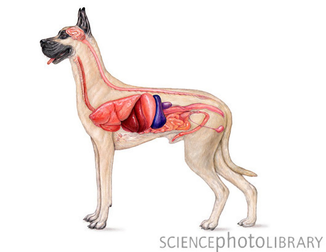
The musculoskeletal system includes all the muscles, bones and joints. The respiratory system ( cat) ( dog) includes the mouth, nose, trachea, lungs and smaller airways (bronchi and bronchioles). The respiratory system is responsible for taking in oxygen and eliminating waste gases like carbon dioxide. Because dogs and cats do not sweat through ...

Dog Skeletal Anatomy Flashcards | Quizlet
Download scientific diagram | ASTM D 638 "dog bone" specimen. from publication: On the Feasibility of Thermoelastic Stress Analysis on Rapid Prototyping Models | This work discusses application of ...
Types and Parts of Bones | Reading Ancient Animal Remains
Speaking of skeletons, a dog has 320 bones in their body (depending on the length of their tail) and around 700 muscles. Muscles attach to bones via tendons. Depending on the breed of dog, they will have different types of muscle fibers. You've probably heard about slow and fast twitch muscle fibers before.

Identify the dog skeleton Quiz
skeleton of a dog. previous. next. caudal vertebrae Bony parts comprising the skeleton of the tail located at the terminal end of the vertebral column. ... Flat skull bone forming the forehead and top of the eye sockets, and articulating especially with the parietal.

Maceration (bone) - Wikipedia
ANIMAL KINGDOM :: CARNIVOROUS MAMMALS :: DOG :: SKELETON OF A ...

Domesticatie Hond en evolutie hondachtigen | Dog skeleton ...
dog - Teeth | Britannica
Skeleton Worksheet Answers - WikiEducator
Pelvis anatomy - The Institute of Canine Biology

Canine Anatomy, Complete Set of 3 Charts. Buy the Set and SAVE!

Dog Anatomy Stock Illustrations – 4,378 Dog Anatomy Stock ...
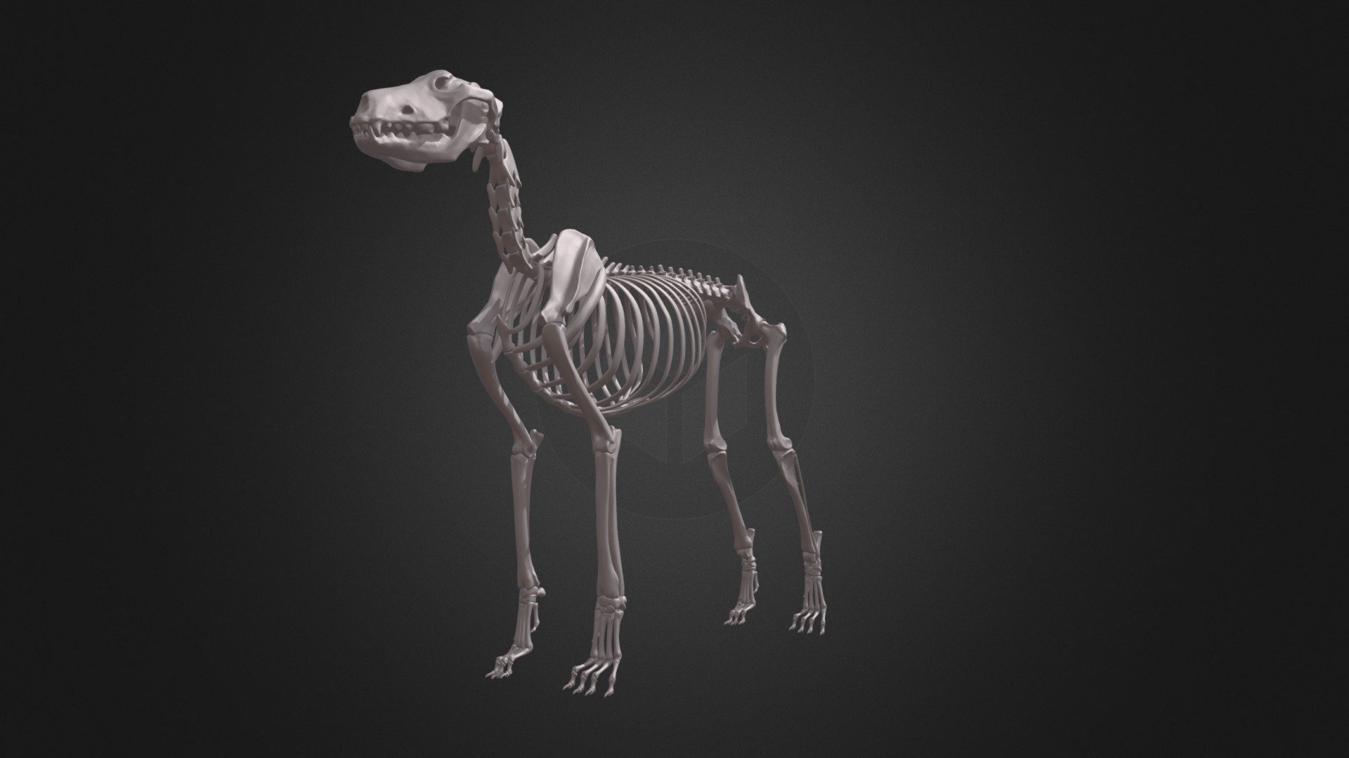
DOG SKELETON - 3D model by zorrenhimself (@zorrenhimself ...
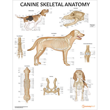
Canine Anatomy - AnatomyStuff Free Resources

Schematic diagram of dog bone shape tensile specimen ...

Complete Ossified Skeleton of a Dog Post Growth Plate Closure ...

skeletal structure of a dog – My Drawing Course

Dog Bone

an atlas of animal anatomy for artists | Dog anatomy, Dog ...
Schematic drawings of the dog-bone test samples. | Download ...

Labeled atlas of anatomy: illustrations of the dog - vet-Anatomy

2,707 Dog Skeleton Stock Photos, Pictures & Royalty-Free ...

a) Direct tensile test setup and (b) geometry of dog bone ...
Dog Skeleton Veterinary Vector Illustration Dog Stock Vector ...

Dog Skeleton Specimen Model Animal Anatomy Teaching Model ...

Schematic of the 4-point conductivity test on dog bone dense ...

Dog bone sample shape and size. | Download Scientific Diagram
Free Dog Anatomy Cliparts, Download Free Dog Anatomy Cliparts ...
Dog Bones labeled by Otvali on DeviantArt
Comments
Post a Comment