39 cranial cavity diagram
Body regions Right or left based on the person being viewed. •A). Superior (cranial) •B). Inferior (caudal) •C). Anterior or ventral •D). Posterior or dorsal •E). Medial •F). Lateral •G). Intermediate •H). Proximal •I). Distal •J). Superficial •K). Deep •L). Peripheral (also means outward) Regional terms •A). Axial: Head neck and trunk •1). In the skull base, there are numerous foramina that transmit cranial nerves, blood vessels and other structures - these are collectively referred to as the cranial foramina.
Oct 30, 2020 · Learn the major cranial bone names and anatomy of the skull using this mnemonic and labeled diagram. Sutures connect cranial bones and facial bones of the skull. Develop a good way to remember the cranial bone markings, types, definition, and names including the frontal bone, occipital bone, parieta
Cranial cavity diagram
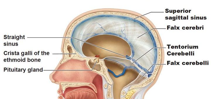
The neurocranium forms the cranial cavity that surrounds and protects the ... This is a diagram of a skull that displays the components of neurocranium. Cranial cavity. Lecture 8 Edited by : Nadeen Al-Falooji. Dr. Heba Kalbouneh. Roofs over the posterior cranial. fossa :Divides the cranial cavity into Ø. SUPRATENTORIAL. Dr. Majid Doroudi guides you through the exploration of the cranial cavity, its contents, and related gross anatomical features.00:00 - Introduction00:16...
Cranial cavity diagram. Oct 24, 2021 · Let’s take a closer look at the layers associated with the cranial cavity. Use the colored diagram in the bottom left of the image as a reference. First, the cranium/skull is on the outside which encloses the cranial cavity. Below the skull is 3 layers of membrane called the meninges. The 3 meningeal layers are labeled with the stars. There are four pairs of sinuses (named for the skull bones in which they're located). Interactive diagrams show sinus cavity locations and help visualize sinusitis, the most common type of sinus ... This bone grows as spicules into the medullary cavity from the endosteal surface, serving as a labile reserve of mineral that can be mobilized to provide calcium for egg shell formation. Scenes from the Past: CT-guided Endoscopic Recovery of a Foreign Object from the Cranial Cavity of an Ancient Egyptian Mummy.
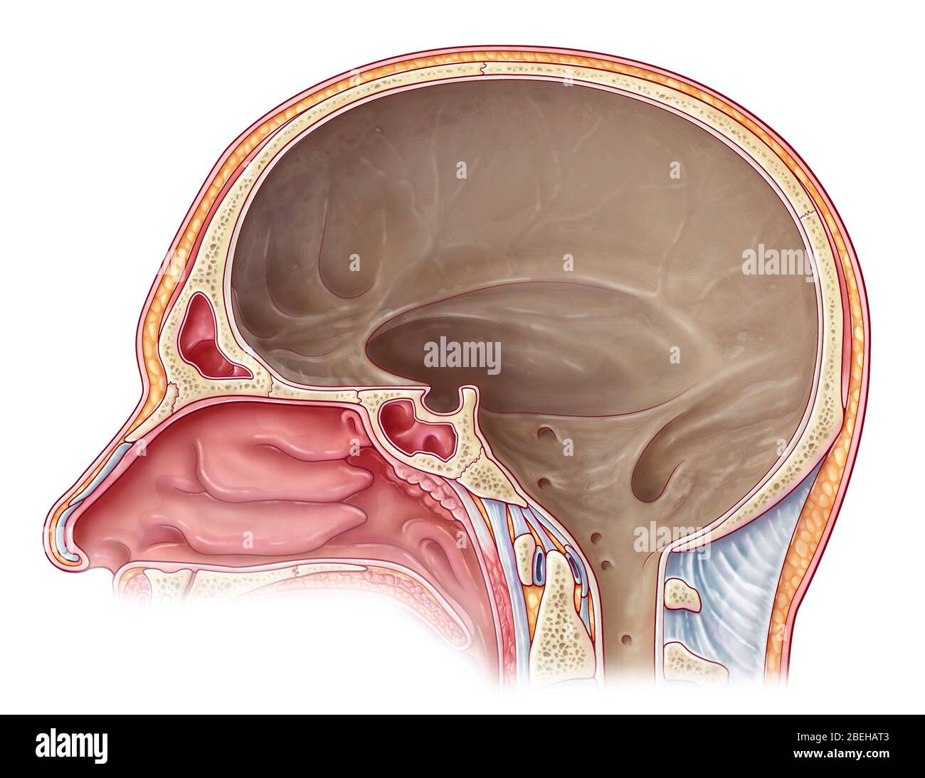
The physics of the cranial cavity, hydrocephalus and normal pressure hydrocephalus: mechanical interpretation and mathematical model. Sep 4, 2021 — The space where the brain is located in the skull is called the cranial cavity. Brain with 4 color coded lobes of cerebral cortex. The image ... This article introduces the cranial nerves, their anatomy, names, functions and mnemonics to help you remember them. Click now to learn more! The cranial cavity, or intracranial space, is the space formed inside the skull. The brain occupies the cranial cavity, which is lined by the meninges and which contains cerebrospinal fluid to cushion blows. Eight fused cranial bones together form the cranial cavity: the frontal, occipital...
Cranial — Cranial. The cranial cavity is the anterior portion of the dorsal cavity consisting of the space inside the skull. This cavity contains the brain, ... introduction to cranial cavity cavity present in cranium of skull is known as cranial cavity. it contains brain , meninges , venous sinuses, all cranial nerves , four petrosal nerve , part of internal carotid artery and a part of vertebral artery besides the special senses. the anterior branch of middle menengial artery lies at the pterion and … Angular measurements and the cranial cavity area were obtained at the mid-sagittal plane To obtain the relative position and growth pattern of jaws to the cranium as well as the relative position of the... Block diagram of numerical solution steps of cranial cavity with the finite element method. Figure 12 is the strain diagrams of cranial cavity under the mild hypothermia environment and normal...
Cranial bones - Nursing Lecture
Cranial Nerves. Evolution. Functions of the Peripheral Nervous System. includes a fluid-filled cavity enclosed within a membrane. A hydrostatic. skeleton usually is further encased within a muscular coat.
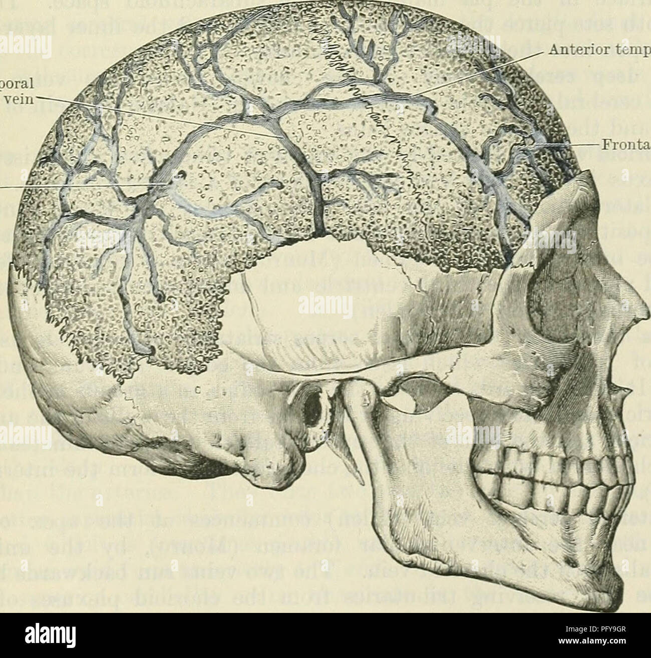
Cunningham's Text-book of anatomy. Anatomy. DIPLOIC AND ...
Schematic diagrams of the bodies of animals with coeloms, with pseudocoeloms, or without body cavities. The cranial cavity cushions and protects the brain within a rigid skull.

Cranial Bones - Comparative Oral+ENT Biology - OpenStax CNX
Identify the cranial foramina and name the structures that pass through them. A - dura Mater B - arachnoid mater C - pia mater.
JaypeeDigital | eBook Reader
The cranial cavity, or intracranial space, is the space formed inside the skull. The brain occupies the cranial cavity, which is lined by the meninges and ...
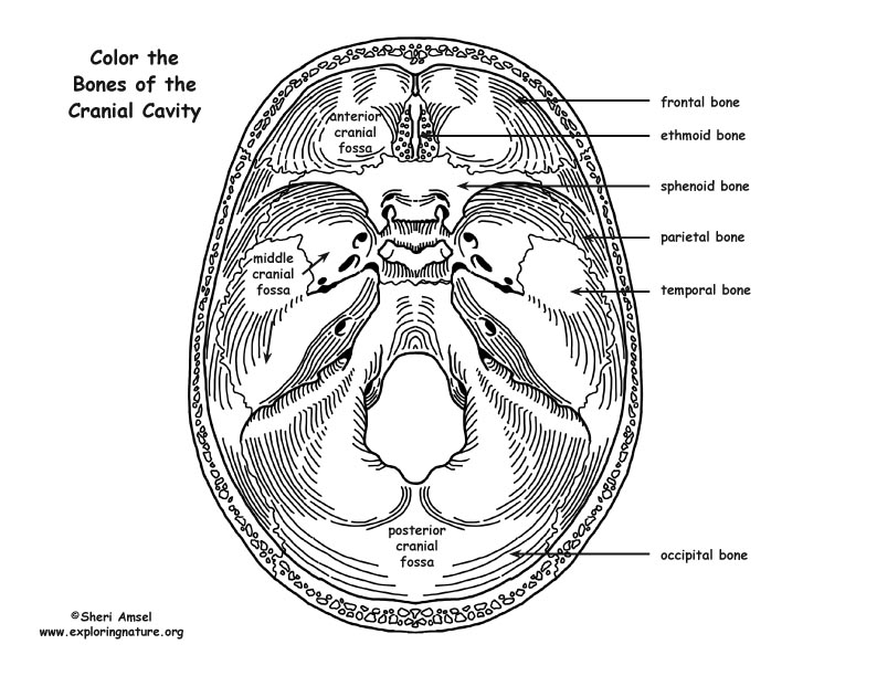
Skull Cranial Cavity Bones Coloring Page
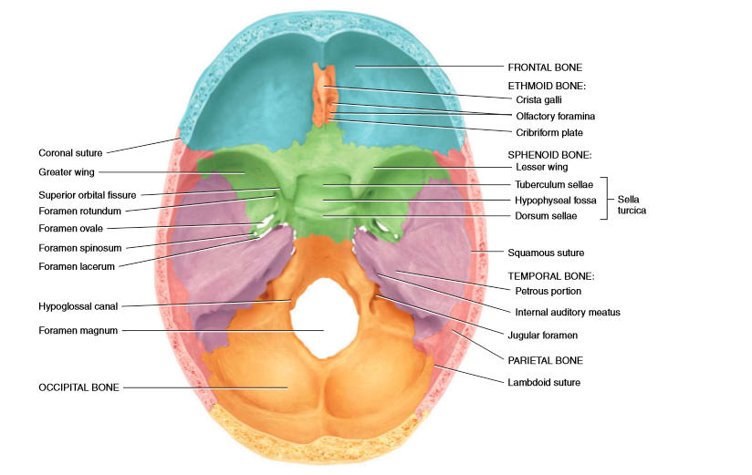
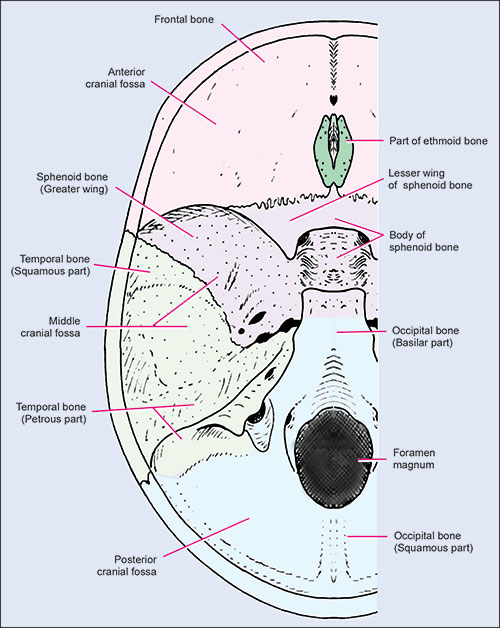
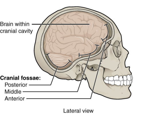
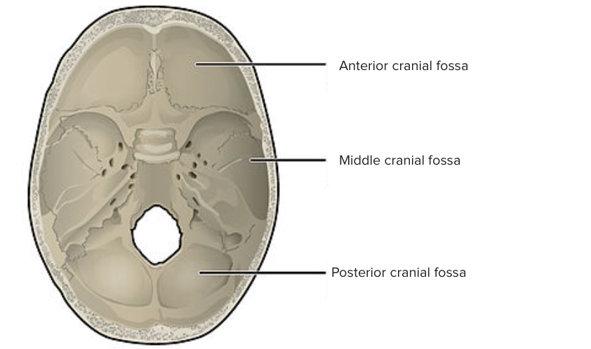
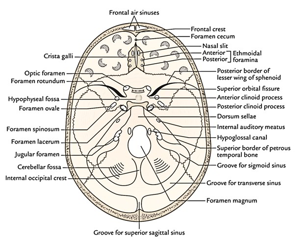
You know there is three-division (rostral, middle, and caudal cranial fossae) present in the base of the cranial cavity. The rostral cranial fossa supports the olfactory bulbs, tracts, and remaining part of the brain’s frontal lobe. It continues rostrally by the concave cribriform plate. Several cribriform foramina perforate this cribriform plate.

Duke Anatomy - Lab 18: Cranial Cavity | Nerve anatomy, Facial ...
Like spinal nerves, cranial nerves are bundles of sensory or motor fibers that innervate muscles or glands; carry impulses from sensory receptors, or show a combination of these fiber types.

Body cavity Human body cavities Cranial cavity Anatomy ...
Nerves which originate from the brain or the brainstem are referred to as the cranial nerves. Do keep in mind that nerves which originate from the spinal cord segments aren't called cranial nerves.

The Skull | Anatomy and Physiology I
31.1 Cranial nerves Inferior (basal) view. The 12 pairs of cranial nerves (CN) are numbered according to the order of their emergence from the brainstem. Note: The sensory and motor fibers of the cranial...
7.2 Head and Neck Basic Concepts – Nursing Skills
Head and Neck The Cranial cavity part II Meninges & Dural Folds Dr. Mohamed Elfiky Professor of anatomy and embryology.
Skull base fracture, posterior
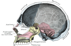
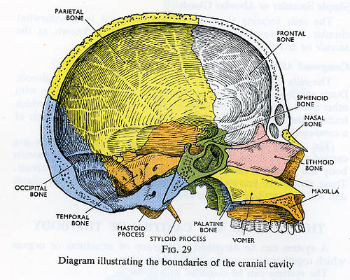
Skull = bones of cranium (enclose cranial cavity) + bones of face (includes the mandible). Notes: calvaria = roof of cranial cavity (intramembranous bones, e.g., frontal, parietal, etc...
Anterior Cranial Fossa - AnatomyZone
Cranial cavity. Quite the same Wikipedia. Just better. As you can see on this diagram, we have the pia mater here which is below the blood vessels and directly on the spinal cord, the arachnoid mater...
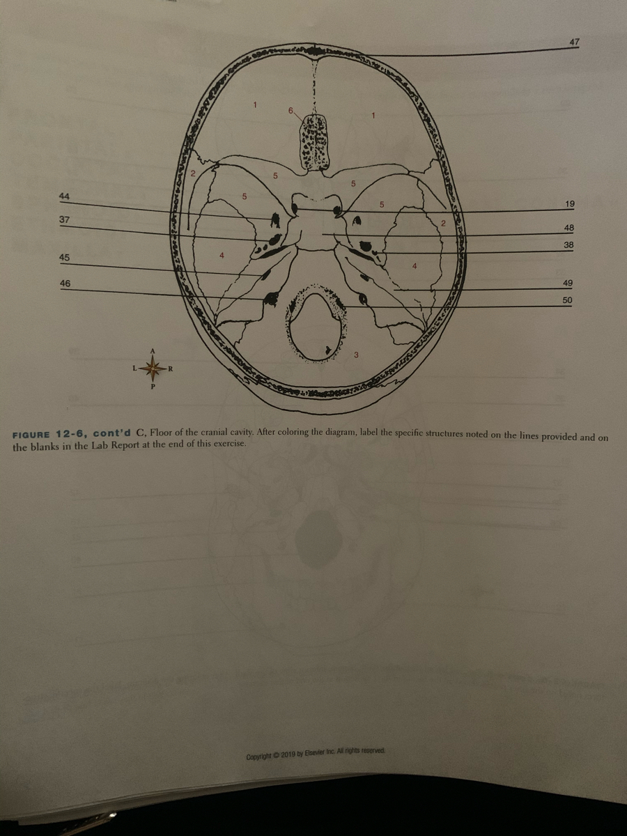
Answered: 44 19 37 2. 48 38 45 46 49 50 FIGURE… | bartleby
bony cavity called orbit (Fig. 1.4). Each eyeball is located in the anterior orbit, nearer to the roof and lateral wall than to the floor and medial wall. Each eye is protected anteriorly by two shutters called...

Intracranial region | Clinical Gate
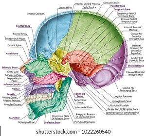
Forms part of middle cranial fossa and lateral aspect of skull (temple) Sphenoid bone Contributes to middle cranial fossa, nasal cavities, orbits, and lateral skull (temples) Contains sella turcica ("Turkish saddle"), sphenoidal air sinus, and many foramina Lesser wing of sphenoid bone

Dissection of cranial cavity showing the infratentorial space ...
The cranial cavity of the horse encloses and protects the brain, its meninges, and vasculature and is formed by the following bones: the frontal, parietal, and interparietal bones create the roof...
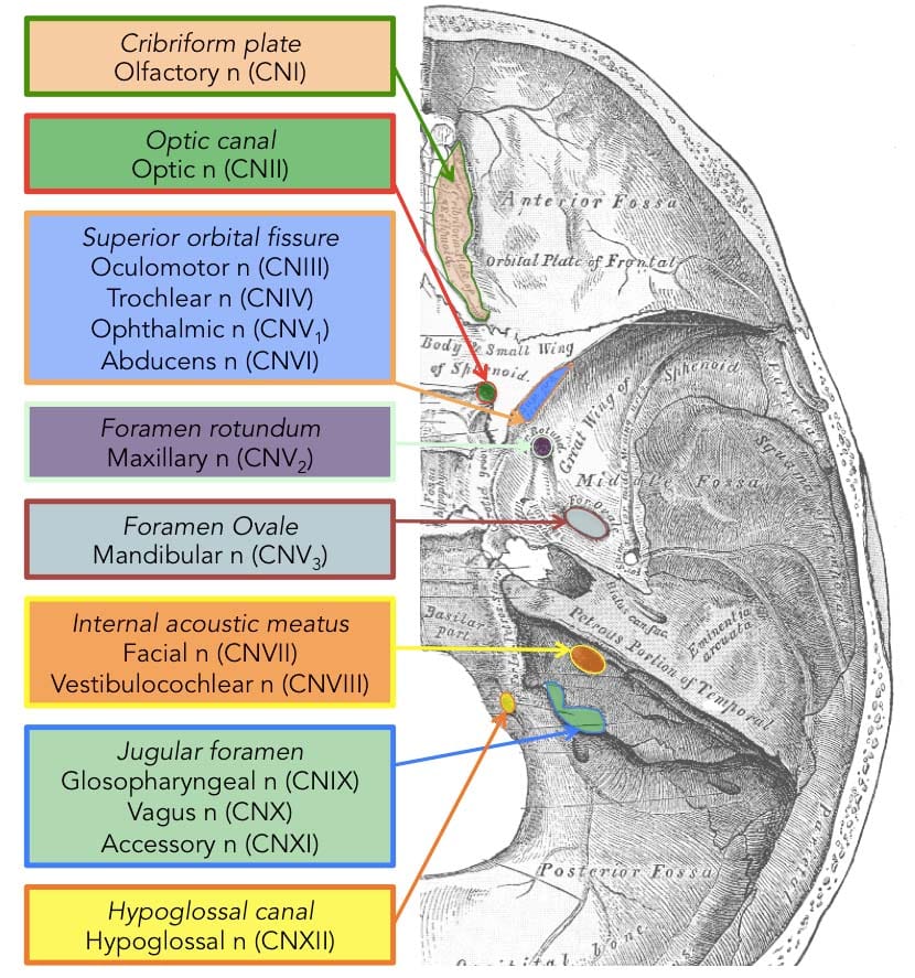
Cranial Foramina - Foramen Ovale - Skull - TeachMeAnatomy
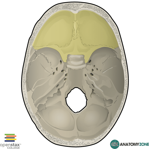
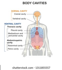
Cavities: - Cranial cavity, - Nasal cavity, - Paranasal sinuses - Oral cavity, - Orbit, - (Tympanic cavity Fossae cranii. Anterior cranial fossa MIddle cranial fossa Posterior cranial fossa.
Cranial cavity Images, Stock Photos & Vectors | Shutterstock
Cranial Cavity is the main cavity of the skull. It lodges the brain, meninges, portions of the cranial nerves and blood vessels. The floor of the cranial cavity is composed by the upper surface of...
Skull | Concise Medical Knowledge
The cranium may be divided into two major areas for study— the cranial vault, or calvaria, forming the superior, lateral, and posterior walls of the skull; and the cranial base, forming.
An illustration of the cranial cavity from a midsagittal view ...
The cranial cavity contains the brain, pineal and hypophysis cerebri, parts of the cranial and spinal nerves, blood vessels, meninges and cerebrospinal fluid. Bones that make up the cranial cavity: Cranial cavity is contained by the frontal, parietal, sphenoid, temporal and occipital bones, and in part the ethmoid, all lines by fibrous endocranium, external zone of dura mater and pericranium.
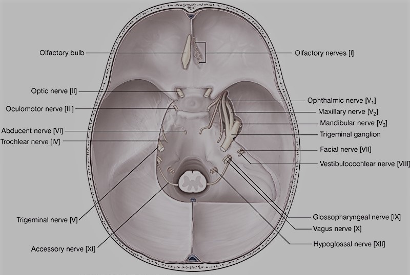
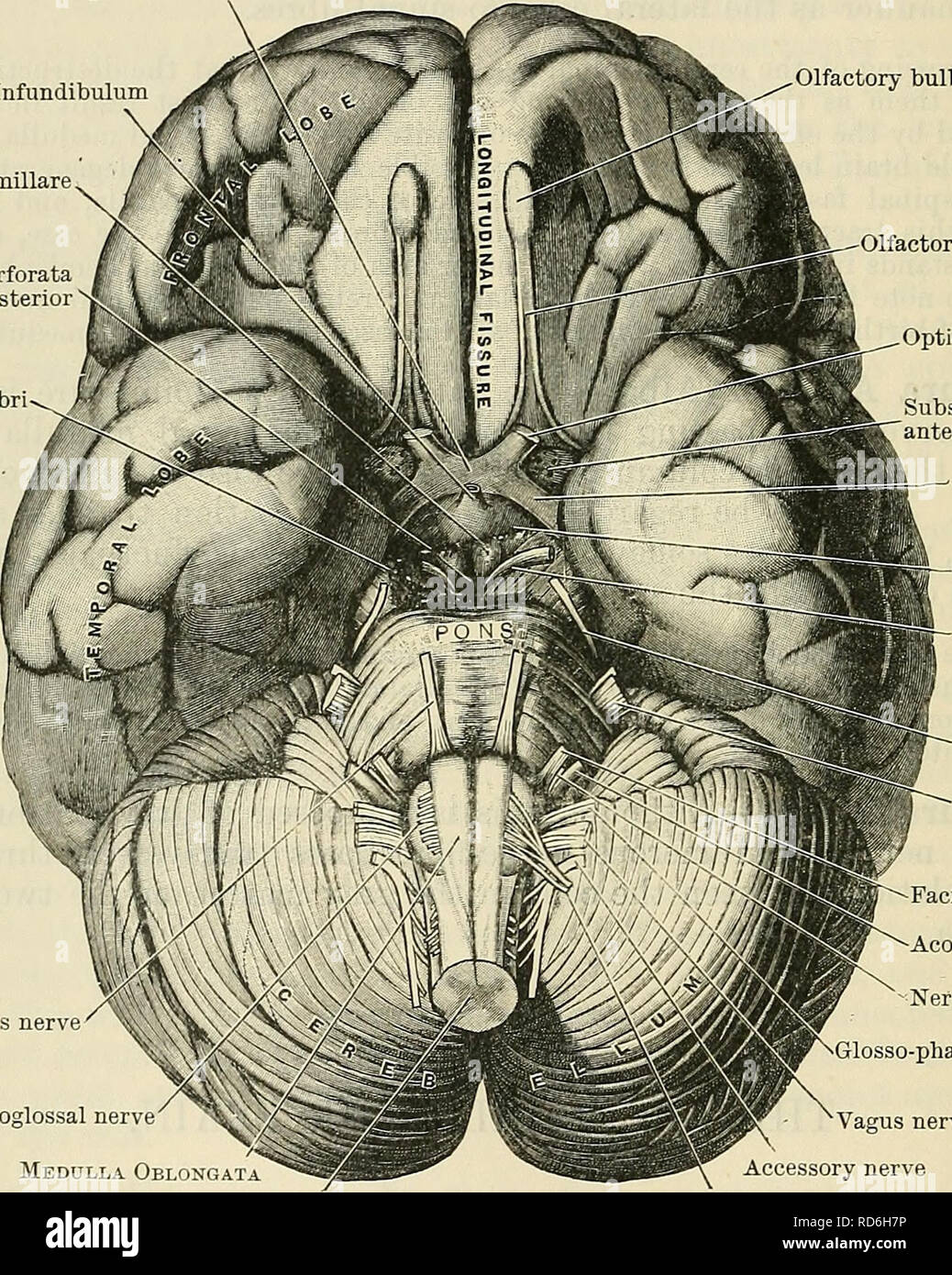
Superior view of the base of the skull: Anatomy | Kenhub
The posterior cranial fossa serves as a bed for the hemispheres of the cerebellum (a mass of brain tissue behind the brain stem and beneath the rear portion of the cerebrum) and for the front and...

the base of the cranial cavity Diagram | Quizlet
Illustrations and diagrams of the 12 pairs of cranial nerves - eAnatomy Anatomical atlas of the cranial nerves: illustrations, anatomical drawings, and diagrams ×Your email address is not verified. Verify now Toggle navigation ANATOMY e-Anatomy vet-Anatomy Anatomy Ninja

Floor of the Cranial Cavity Diagram | Quizlet
The cranial cavity, also known as intracranial space, is the space within the skull that accommodates the brain. The skull minus the mandible is called the cranium. The cavity is formed by eight cranial bones known as the neurocranium that in humans includes the skull cap and forms the protective...
Cranial cavity - Wikipedia
The cranial nerves also control balance, hearing, and swallowing. The twelve cranial nerves, in order from I to XII are: olfactory nerve, optic nerve, oculomotor nerve, trochlear nerve, trigeminal nerve...
Cranial Cavity
Explore the interactive 3-D diagram below to learn more about the cranial bones. Cranial bone conditions Several injuries and health conditions can impact your cranial bones, including fractures...

Cranial cavity Images, Stock Photos & Vectors | Shutterstock
For Sensory System Modula lecture skull and cranial cavity skull all bones of the skull are attached - Mandible is not part of the cranium or facial skeleton Cranium can be divided into: - Calvaria the...

Cranial Cavity Bones Cranium Bones Head Stock Illustration ...
Dr. Majid Doroudi guides you through the exploration of the cranial cavity, its contents, and related gross anatomical features.00:00 - Introduction00:16...

ImageQuiz: Skull - Cranial Cavity FINAL
Cranial cavity. Lecture 8 Edited by : Nadeen Al-Falooji. Dr. Heba Kalbouneh. Roofs over the posterior cranial. fossa :Divides the cranial cavity into Ø. SUPRATENTORIAL.

Head & Neck, Lesson 2
The neurocranium forms the cranial cavity that surrounds and protects the ... This is a diagram of a skull that displays the components of neurocranium.
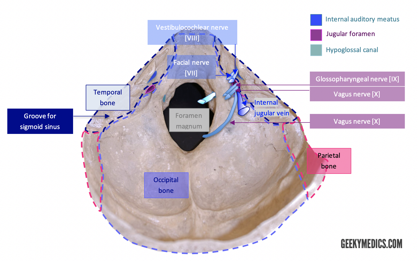
Cranial Foramina | Skull Anatomy | Foramen | Geeky Medics

Cranial Bones - Comparative Oral+ENT Biology - OpenStax CNX
Cunningham's Text-book of anatomy. Anatomy. 540 THE NEKVOUS ...
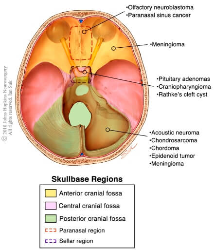
Types of Skull Base Tumors and Conditions | Johns Hopkins ...
Cranial Cavity – Earth's Lab

What is the Cranial Cavity? (with pictures)
cranial cavity function | Science online

Anterior Cranial Fossa - ppt download
What is the cavity where the brain is located? | Socratic

Cranial fossa - Wikipedia

Bones of the Cranium - Course Hero
Cranial cavity Images, Stock Photos & Vectors | Shutterstock
Comments
Post a Comment