41 cow bones diagram
Clavicle*diagram* Clavicle* Human*Ulnaand*Radius* Human*humerus* Human*humerus*and*scapula. Human*Humerus*and*Scapula. Human*sacrum* ... Human*vertebrae* Skull*and*Mandible* Inside*of*skull* Child. Infant. Mummified*bear*vs.*Human* Bear*on*leV* Side View of Foot Bones Inter mediate gone gone gone Talus gone Ca can eug gone Cuboid gone gores ... Cow Leg Bone Structure. Bull soundness structural ot 4235 cow femur diagram avian skeletal system small and forward facing front knees like horses general anatomy of the bull and cow. Lab 5 cow horse leg foot muscle and skeleton anatomy the skeletal system of a cow by tony smith 28 bones diagram bone keywords the skeletal system of a cow by ...
Cattle and horse long bones also show very distinct differences especially (but not exclusively) femora and metapodials. Figure 3 shows cattle and horse femora. The most apparent difference in these bones is the much larger muscle attachments at the proximal end of the horse femur compared to cattle (horses run faster than cattle!).

Cow bones diagram
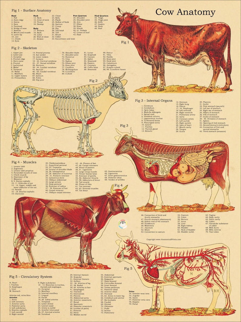
THE NECK. The vertebral column/backbone is the main axis of the skeleton and it protects the spinal cord. The spinal cord is located in a neural canal formed by a long series of neural arches. The neural arch of each vertebra is supported on the body or centrum of the vertebra. In types of vertebrae,the neural arch extends as a prominent spine ... Chapter 4 Cow Anatomy Terms to Know Pins- the protrusions of the wing of the Ilium bones (pelvis) just lateral to the base of the tail in ruminants Brisket- the mass ... - A free PowerPoint PPT presentation (displayed as a Flash slide show) on PowerShow.com - id: 494b88-ZjdiY Pelvic Bone Oviducts Cervix Ovaries Teats . Figure 3: Cow silhouette showing location of the reproductive tract . Vulva . The outermost portion of the reproductive tract is the vulva. This structure protects the reproductive tract by keeping out larger debris. Although the vulva is a relatively small portion of the entire tract, its visibility ...
Cow bones diagram. PDCA - One Blog. Welcome to the first Dexter cattle blog to disseminate information for members of the Purebred Dexter Cattle Association of North America (PDCA) and for those with a curiosity about Irish Dexter cattle, cattle in general, as well as news from the PDCA. Expressions of opinion are to not be regarded as expressing the official ... Browse 928 cow anatomy stock photos and images available, or search for pancreas or bovine anatomy to find more great stock photos and pictures. American Meat cuts diagram poster design. Beef scheme for butcher shop vector illustration. Cow animal silhouette vintage retro hand drawn style graphic. Bovine Bone Specifics. Ilium. In the cow the tuber coxae is visible and is readily palpable. The sacral tuber has two prominences; the cranial and caudal dorsal iliac spines. The iliac crest is thin and concave. The ileal wing is orientated in a vertical manner. Ischium. The ischial tuberosity is triangular in shape. Femur. Animal bones have several uses to a witch. Probably primarily they are used for divination - the layout and positioning of the bones can be read in the same way as a tarot or a rune spread. The bones can also be used in spell casting to draw upon the energies of the animal and to invoke its spirit into the workings.
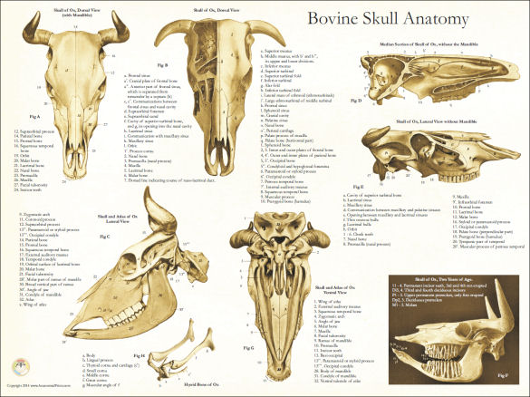
The maxilla bone of a pig is the main bone of the upper jaw and carries upper check teeth. Palatine bone locates on either side of the caudal nares of a pig. In the rostral margin of the bony orbit, there is an irregular lacrimal bone in the pig skull. Now, I will show you the different bones with a diagram from the pig skeleton skull anatomy. T-Bone Steak Porterhouse Steak o DI DI, D2, D3 Boneless Sirloin Steak DI Pin Bone Sirloin D2 Flat Bone Sirloin D3 Wedge Bone Sirloin El Boneless Rump Roast E2 Top Round Roast Small End Rib Steak, Small End LOIN Top Loin Steak Boneless T-Bone Steak Tenderloin Roast (Filet Mignon) Porterhouse Steak Tenderloin Steak (Filet Mignon) RIB 9.50/0 SIRLOIN Where does the T-bone come from on the cow? short loin The T-Bone is cut from the short loin, and actually has two different steaks attached to the bone. On the long side is the strip. If you would take that strip and cut it away from the bone, you would have Rube's New York Strip. On the smaller side of the T-bone is the tenderloin. The diagram shows the way in which the food passes into the rumen, circulates to the reticulum, and is later regurgitated into the mouth for further chewing. When swallowed a second time some of the food returns to the rumen while the remainder passes through the omasum into the abomasums and thence into the small intestine.
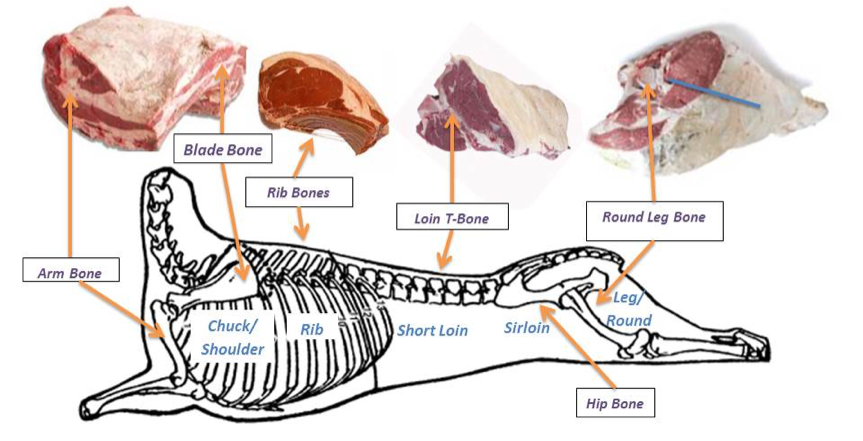
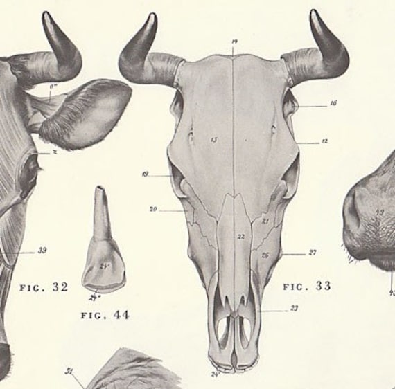
4. Loin. Sub-Primal Cuts: Porterhouse, T-bone, Club Steak, Filet Mignon, New York Strip, Sirloin Steak, Sirloin Cap, Chateaubriand, Tri-Tip. Location: Located on the lower back of the cow, the loin is a less active muscle that runs along the spine and tends to be tender and soft making it ideal for juicy steaks. Ingredients Beef Cuts Chart and Diagram, with Photos, Names, Recipes, and More. Learn all about the most popular beef cuts from our chart, diagram and write up, including popular and alternative names, where the cuts come from on the cow, preferred outdoor cooking methods, their costs relative to each other, and a fantastic recipe for each cut of beef that we've taken from around the web. Atlas of bovine anatomy: the essentials of the bull and the cow (skeleton, joints, muscles, parts and region of the body) ... (skeleton, bones, muscles, joints and viscera). Positional and directional terms are also illustrated. The drawings and anatomical labeling of these illustrations were done by Gauthier Kervyn, under the anatomical and scientific supervision of Antoine Micheau - MD and ... The cow has 6 phalanges (three in each toe). For comparison, humans have 26 foot bones, comprising 7 tarsals, 5 metatarsals (one leading to each toe) and 14 phalanges (two for the big toe and three for every other toe). 15-21 are the ankle bones, 23 and 24 are the metatarsals, and 26-28 show the three phalanges in each toe.
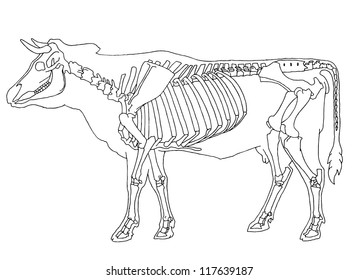
Cow Skeleton Diagram Labeled - Diagram Media
Cow Bone Structure. December 14, 2017 - by Wandi - Leave a Comment. Skeletal structure general anatomy of the bull and cow meat fabrication methods the dz 8588 cow femur diagram schematic wiring. General Anatomy Of The Bull And Cow Ilrated Atlas. The Skeletal System Of A Cow By Tony Smith.

Gluten Intolerance, bone loss, and nutritional deficiencies.mp4
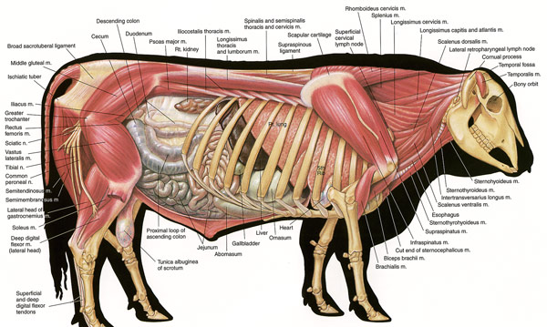
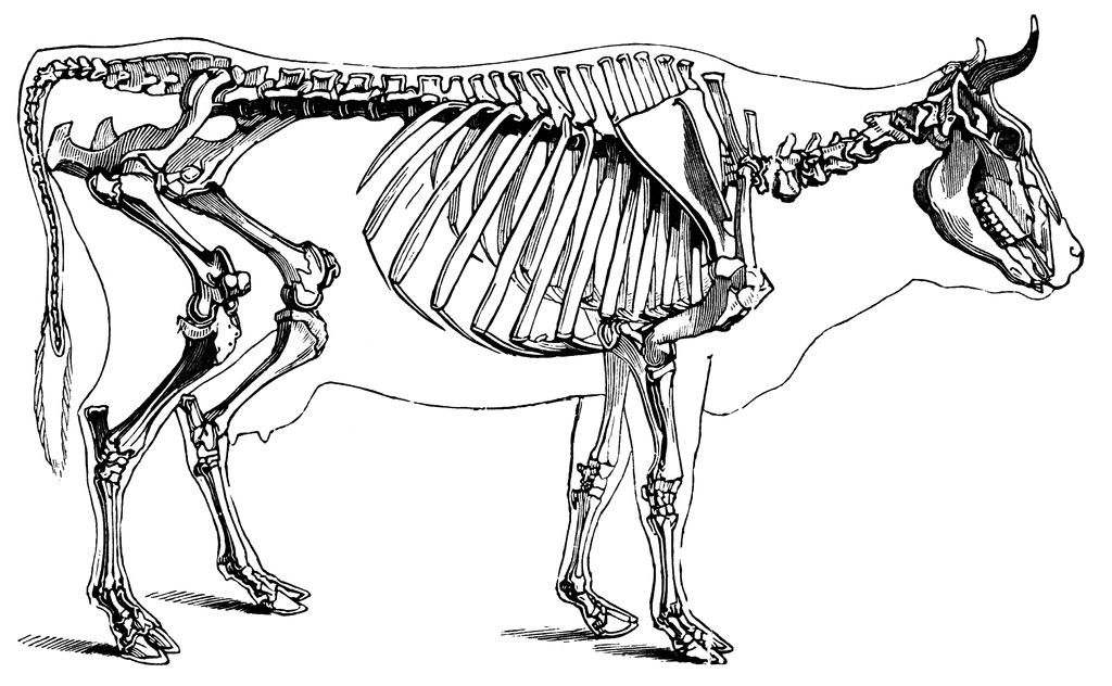
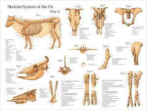
The bones of the forelimb of a cow – scapula, humerus, radius-ulna, carpal, metacarpal, and phalanges. Hindlimb bones of a cow – include ilium, ischium, pubis, femur, tibia-fibula, tarsal, metatarsal, and phalanges. The bones from an axial skeleton of a cow – include bones of the skull, vertebrae, ribs, and sternum.

Special Problems. Carving Meat
cow bone lab. STUDY. Learn. Flashcards. Write. Spell. Test. PLAY. Match. Gravity. Created by. Ethan_Brick PLUS. Terms in this set (8) Epiphysis. At the end of the bone. The articular surface is covered with hyaline cartilage. Epiphysis. At the end of the bone. The articular surface is covered with hyaline cartilage.

Cow - legs | Anatomy | Pinterest | Cattle, Cow and Anatomy
Cow Anatomy - Diagrams Of Cows & Calves Cow Anatomy Below is a diagram of the Anatomy of a Cow As you can see, there are many parts to a cow. Cows vary in all different colours, some are brown, tanned, white, black, brown-white patched or black-white patched. In a female cow, milk is produced in the udders and extracted from the teats.

Cuts of beef | ClipArt ETC
You may also find cow skeleton, cow internal organ, cow bone, cow stomach, cow milk as well. The biological subfamily Bovinae includes a diverse group of 10 genera of medium to large-sized ungulates, including domestic cattle, bison, African buffalo, the water buffalo, and the four-horned and spiral-horned antelopes. The evolutionary relationship between the members of the group is still ...

I made this picture at the wildness in Austria, phontanella. I’ve won the prize of National Geographic junior in 2015.
The T in the T bone is named because the spinal processes are at roughly 90∘ to the vertebrae. This diagram may help: Taken from the nose to tail app. Typically a t bone is quite far down the cow, as shown here as the short loin: Taken from Wikipedia, beef cut.

Cow Skeleton Diagram Printable Digital Graphic Download ...
An adult animal weighs on average about a ton, so the skeletal bones of a cow are large and durable. They form a robust frame that can withstand a significant load. The spine is the axis to which the skull, ribs, shoulder blades, pelvic bones, and the tail of the cow are attached. The limbs are attached to the shoulder blades and pelvis.

Pin on logic
Heterotopic bones — os penis [ carnivore; rodent ] os cardis [ cattle ] Shape: Long bones — length greater than diameter Short bones — approximately equivalent dimensions Flat bones — e.g., scapula, os coxae, many bones of skull Irregular bones — short & multiple processes (vertebrae) Sesamoid bones — small "seed-like" within ...

Image from page 31 of "Agriculture .." (1901)
Here's our Cow Map, a diagram of all the key sections of a cow and what cuts of beef come from each area. Key Sections of a Cow . Here are the major sections of a butchered cow along with the cuts of beef that come from them. Brisket - brisket flat cut; Chuck - boneless short ribs, shoulder petite tender medallions, shoulder petite tender, boneless shoulder pot roast, boneless shoulder steak ...

Cow Leg Bones Diagram - Diagram Media
Download 499 Sheep Diagram Stock Illustrations, Vectors & Clipart for FREE or amazingly low rates! New users enjoy 60% OFF. 177,954,846 stock photos online.
cattle
Metacarpal bones. These are covered in detail in the bovine lower limb section. Joints of the Distal Forelimb Carpal Joint. The carpal joint is a compound joint composed of: 1. The antebrachiocarpal joint between the radius/ulna and the proximal carpal bones. 2. The middle carpal joint between the two rows of carpal bones. 3.
Vintage Anatomical Chart of the Cow 24 X 36
Pelvic Bone Oviducts Cervix Ovaries Teats . Figure 3: Cow silhouette showing location of the reproductive tract . Vulva . The outermost portion of the reproductive tract is the vulva. This structure protects the reproductive tract by keeping out larger debris. Although the vulva is a relatively small portion of the entire tract, its visibility ...

Cow Leg Bones Diagram / Human Skeleton Labeled Diagram ...
Chapter 4 Cow Anatomy Terms to Know Pins- the protrusions of the wing of the Ilium bones (pelvis) just lateral to the base of the tail in ruminants Brisket- the mass ... - A free PowerPoint PPT presentation (displayed as a Flash slide show) on PowerShow.com - id: 494b88-ZjdiY
anatomy of the cow
THE NECK. The vertebral column/backbone is the main axis of the skeleton and it protects the spinal cord. The spinal cord is located in a neural canal formed by a long series of neural arches. The neural arch of each vertebra is supported on the body or centrum of the vertebra. In types of vertebrae,the neural arch extends as a prominent spine ...

Image from page 177 of "Bulletin : report of Agricultural Experiment Station, Agricultural and Mechanical College, Auburn, Ala" ([1888-1903])

Cow Skull Anatomy Poster - 18" X 24" | mandible ...
Vintage Anatomical Chart of the Cow 24 X 36

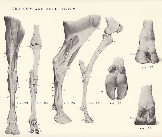
The femur in camel, cow and mare (from left to right): A ...
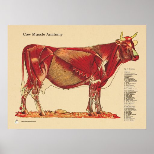
Cow Bovine Veterinary Muscles Anatomy Chart Poster | Zazzle

Cow Leg Bones Diagram - The femur in camel, cow and mare ...
Cow Skeleton | ClipArt ETC

Cow Leg Bones Diagram / General Anatomy Of The Bull And ...

Bovine Anatomy - Skeletal … | Cow anatomy, Large animal ...

Sunset views from my rooftop

Canadian Beef Butchering cuts...Diagram and Bone Structure ...
Bull Anatomy
Meat Fabrication Methods — The Culinary Pro
Set of 2 Vintage Illustrated COW and BULL Anatomy Skeleton
Cow Anatomy Posters
Bovine Skull Anatomy Poster

Skeleton of a cow showing the anatomical position of the ...
Cow Skeletal Internal Anatomy Poster 18 X 24 | Etsy

cow diagram anatomy... | Cow skeleton, Anatomy, Cow ...

Skeleton of a cow showing the anatomical position of the ...
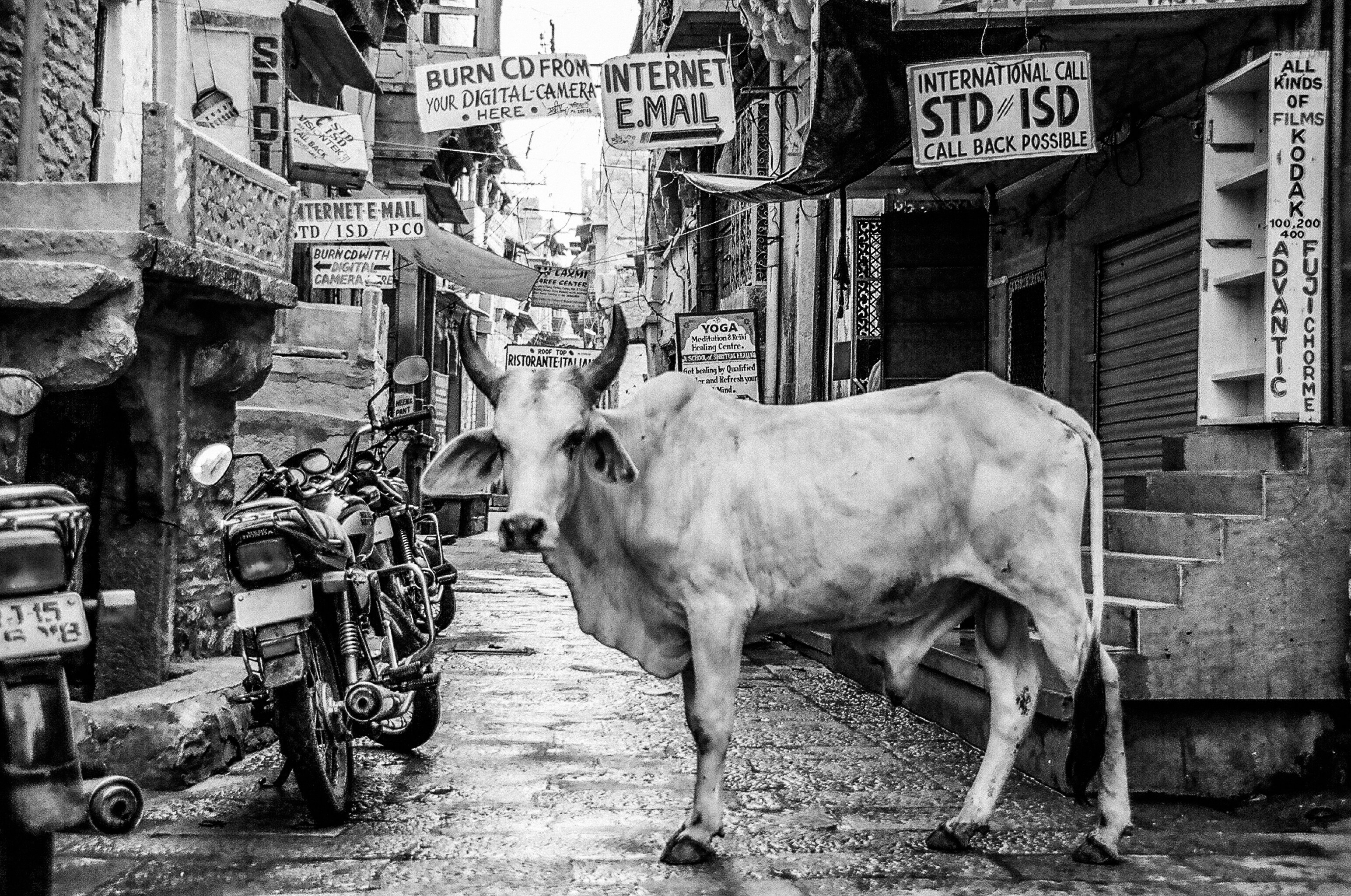
monochrome bull in alley

Cow Leg Bones Diagram / General Anatomy Of The Bull And ...

Cow Skeleton Parts - All About Cow Photos

Image from page 302 of "The American farmer's encyclopedia and dictionary of rural affairs : embracing all the most recent discoveries in agricultural chemistry" (1844)
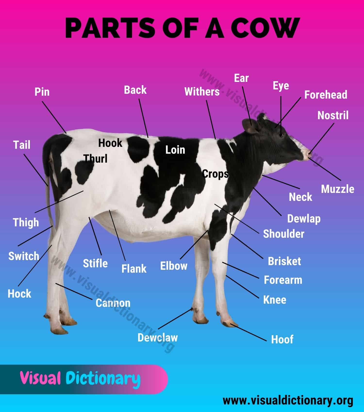
Cow Anatomy: 35 Different External Parts of a Cow (with ...
Items similar to Vintage Cow and Bull Leg and Hoof ...

Image from page 94 of "The diseases of live stock and their most efficient remedies : including horses, cattle, cows, sheep, swine, fowls, dogs, etc. ..." (1886)
Comments
Post a Comment