40 bone cross section diagram
bone cross section diagram chart bone bones bone diagram bone chart anatomy biology science. All Products. bone cross section diagram chart bone bones bone diagram bone chart anatomy biology science. Other Info. Product ID: 239545777421064119Created on: 4/24/2013, 10:27 AM. Cross-Section Diagram Of Human Body - Bones and Organs. Cross section human tendon under light microscope view for education histology. Human tissue, Dense regular connective tissue. Haematoxylin and eosin staining. Cross section of spinal cord under the microscope view. Histological for human physiology
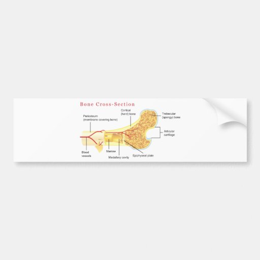
Looking at a bone in cross section, there are several distinct layered regions that make up a bone. The outside of a bone is covered in a thin layer of dense irregular connective tissue called the periosteum. The periosteum contains many strong collagen fibers that are used to firmly anchor tendons and muscles to the bone for movement.

Bone cross section diagram
upper right femur leg bone - bone cross section stock illustrations. raw pork loin chops shot from above on dark background - bone cross section stock pictures, royalty-free photos & images. human bone, cross section diagram of femur showing osteon, veins, marrow. - bone cross section stock illustrations. Vector Illustration Scheme Of Bone Cross Section. Diagram With Articular Cartilage, Marrow, Spongy Bone, Medullary Cavity, Endosteum, Diaphysis, And Periosteum. Bone Cross . Cross Section Of The Spinal Cord. Tooth Section Medical Illustration. Diagram Of Human Bone Anatomy. Cross section through the tongue and C2: Diagram This cross-section has the exact same orientation as the previous one. The posterior landmark is provided by the second cervical vertebra (axis) while the anterior one is provided by the tongue. However, there are quite a few differences between them.
Bone cross section diagram. The best selection of Royalty Free Skin Cross Section Diagram Vector Art, Graphics and Stock Illustrations. Download 100+ Royalty Free Skin Cross Section Diagram Vector Images. Medical diagram. bone marrow cross section stock illustrations. Femur Bone Structure Femur bone structure. Human health concept useful for medical, anatomy and biology educational poster design. Vector illustration with detailed information isolated on a white background. bone marrow cross section stock illustrations. The structure of a long bone consists of several sections:. Diaphysis: This is the long central shaft. Epiphysis: Forms the larger rounded ends of long bones. Metaphysis: Area between the diaphysis and epiphysis at both ends of the bone. Epiphyseal Plates: Plates of cartilage, also known as growth plates which allow the long bones to grow during childhood. . Once we stop growing (between 18 ... I don't like way you've shown the cartilage. It seems confusing and misleading. For example, to read this diagram literally, since the cartilage can be seen inside the cutaway section of bone, it incorrectly indicates that the cartilage in fact goes through the bone structure, rather than just being found around the bone end.
Vector illustration scheme of bone cross section. Diagram with articular cartilage, marrow, spongy bone, medullary cavity, endosteum, diaphysis, and periosteum. Explaned distal and proximal epiphysis. Osteoporosis, Symptoms, Health Care Concept. Vector illustration scheme of bone cross section. Diagram with articular cartilage, marrow, spongy bone, medullary cavity, endosteum, diaphysis, and periosteum. Explaned distal and proximal epiphysis. Bone spongy structure Bone spongy structure vector illustration, normal and with osteoporosis Cross Section. The bladder, like the stomach, is an expandable sac that contracts with inner folds when it is empty. The inner lining of the bladder tucks into the folds and expand out to ... Vector illustration scheme of bone cross section. Diagram with articular cartilage, marrow, medullary cavity and periosteum.. Illustration about educational, diaphysis - 120316017
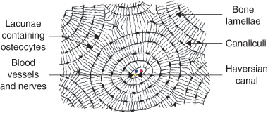
Download scientific diagram | Structure of compact bone. Longitudinal and cross-sectional view of Haversian system in the diaphysis of a long bone shows osteocytes embedded with mineralised ... Cross-section of the Long Bone. Create healthcare diagrams like this example called Cross-section of the Long Bone in minutes with SmartDraw. SmartDraw includes 1000s of professional healthcare and anatomy chart templates that you can modify and make your own. Diagram of Compact Bone (a) This cross-sectional view of compact bone shows the basic structural unit, the osteon. (b) In this micrograph of the osteon, you can clearly see the concentric lamellae and central canals. LM × 40. (Micrograph provided by the Regents of University of Michigan Medical School © 2012) Teeth cross-section, artwork. The upper (biting) surfaces of the tooth are at top, with the lower sections (bottom) embedded in the gums and jaw bone (not shown). The cross-section shows the tooth's internal anatomy, including the living tissue of the pul ID: F6YBB8 (RF) Osteons of a compact bone (cross section) under the microscope.
(a) Layout of bone marrow in a cross-sectional view of a ...
A cross section of a human long bone. The drawings were done with felt markers on a white board in. 3d render of tooth in gums with gutta percha over white background. Here, we basically have a cross section of a piece of bone. Whereas a long bone has only one layer of compact bone (see fig 1). This is known as the periosteum.

Paper cut cross laying on a paper background
Cross-sectional area: The cortical bone equivalent area of the cross-section of the region of interest (femoral neck or shaft), with all soft tissue voids (trabecular and cellular spaces) eliminated (cm 2). Cross-sectional area is derived from the integral of the bone mass profile across the narrow region.

The bones and types of bone cancer - Cancer information ...
53 Bone Marrow Cross Section Premium High Res Photos. Browse 53 bone marrow cross section stock photos and images available, or search for bone cross section or bone cells to find more great stock photos and pictures. human bone, cross section diagram of femur showing osteon, veins, marrow. - bone marrow cross section stock illustrations.

Bone Gallery
Download scientific diagram | Schematic diagram of long bone cross section [47]. from publication: Development of biomimetic electrospun polymeric biomaterials for bone tissue engineering. A ...
Cross Section Through Lower Third of Thigh | ClipArt ETC
Find the perfect cross section bone stock photo. Huge collection, amazing choice, 100+ million high quality, affordable RF and RM images. No need to register, buy now!

1: Schematic drawing of a longitudinal section through a ...
Cross-section. The chambers of the heart operate as a 'double-pump' system for the body's circulation. In coordination with valves, the chambers work to keep blood flowing in the proper ...

Cross Section of Right Kidney - Stock Image - F031/6574 ...
Figure 6.3.6 - Diagram of Compact Bone: (a) This cross-sectional view of compact bone shows several osteons, the basic structural unit of compact bone. (b) In this micrograph of the osteon, you can see the concentric lamellae around the central canals. LM × 40. (Micrograph provided by the Regents of University of Michigan Medical School © 2012)

The Body of Light in the Western Esoteric Tradition...The Middle Pillar...The name of the exercise is taken from the position of the central Sephiroth on the diagrammatic Tree of Life.
Download for free. Royalty-free stock vector ID: 1125379130. Vector illustration scheme of bone cross section. Diagram with articular cartilage, marrow, spongy bone, medullary cavity, endosteum, diaphysis, and periosteum. Explaned distal and proximal epiphysis.

king cross roof architecture
Diagram of Compact Bone (a) This cross-sectional view of compact bone shows the basic structural unit, the osteon. (b) In this micrograph of the osteon, you can clearly see the concentric lamellae and central canals. LM × 40. (Micrograph provided by the Regents of University of Michigan Medical School © 2012)

BBC - GCSE Bitesize Science - Endoskeletons and ...
Second illustrations is a cross section of a 3-pane window labeling all the different parts of the window including the frame. When you're replacing windows, it's nice to know all the different parts of the window and window frame. That's here these 2 diagrams come into play. The first illustrates the anatomy of a window and frame.

Human Skeleton, Natural History Museum, Geneva, Swiss Confederation.
Description: Vector illustration scheme of bone cross section. Diagram with articular cartilage, marrow, spongy bone, medullary cavity, endosteum, diaphysis, and periosteum. Explaned distal and proximal epiphysis.
Bone Cross Section Diagram Car Bumper Sticker | Zazzle
Cross section through the tongue and C2: Diagram This cross-section has the exact same orientation as the previous one. The posterior landmark is provided by the second cervical vertebra (axis) while the anterior one is provided by the tongue. However, there are quite a few differences between them.
Intentionality
Vector Illustration Scheme Of Bone Cross Section. Diagram With Articular Cartilage, Marrow, Spongy Bone, Medullary Cavity, Endosteum, Diaphysis, And Periosteum. Bone Cross . Cross Section Of The Spinal Cord. Tooth Section Medical Illustration. Diagram Of Human Bone Anatomy.

The 100 Best Car Blogs of 2017
upper right femur leg bone - bone cross section stock illustrations. raw pork loin chops shot from above on dark background - bone cross section stock pictures, royalty-free photos & images. human bone, cross section diagram of femur showing osteon, veins, marrow. - bone cross section stock illustrations.

Vector illustration scheme of bone cross section. Diagram ...

Dupuytren Anatomy | Dupuytren Research Group

Structure of compact bone. (a) Cross-sectional view of ...

Hiking in autumn
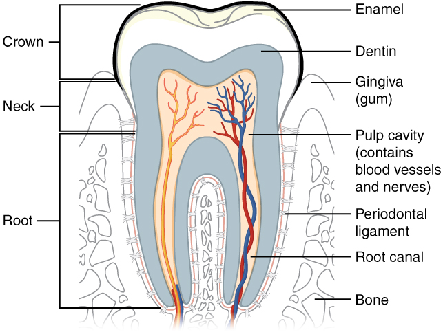
The Mouth, Pharynx, and Esophagus · Anatomy and Physiology

Image from page 243 of "Babyhood" (1886)

Diagram of a cross section of the coiled cochlea ...

Cross and sunset

Schema depicting a cross-section of skeletal muscle fibers ...

cross_section_diagram_of_a_general_human_bone_poster ...

Cross section 6

Gallstones Anatomical Cross Section Vector Illustration ...

Problem Solving Using the Why Tree Video

Knee Joint Cross Section - Medical Art Library

I love the buzz and vibe you get from living in London. I really love the muted colours in this shot and wonder what the guy is actually doing, maybe I should have stopped to have a conversation. Nevertheless, I just had to capture the moment.

Miocene epoch, Florida dugong Rib Bone – Prehistoric Florida

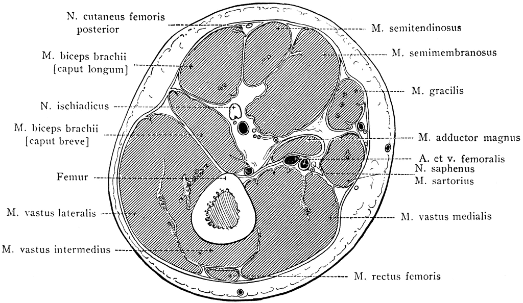
Image from page 692 of "A treatise on orthopedic surgery" (1910)

Studying the composition of bones. This is an osteon from ...

CSA Images 769907

Ontario Heritage Act, Flower, Casa Loma Gardens, Toronto, Ontario, Canada,

3D Bone Marrow Made from Silk Biomaterials Successfully ...
1 Oral embryology, histology and anatomy | Pocket Dentistry

Continuous RANKL Inhibition in Osteoprotegerin Transgenic ...

Dakota Hogback (west of Denver, Colorado, USA) 2

Figure 1-11. Longitudinal section of a long bone (femur ...

Spinal cord sections scheme and vertebra cross section ...
Comments
Post a Comment