39 nerves of the foot and ankle diagram
Symptoms include ankle pain radiating into the foot which tends to be aggravated by walking. Examination may reveal Tinel's sign (radiating pain following nerve percussion) over the tibial nerve at the ankle, weakness and atrophy of the small foot muscles, or loss of sensation in the foot. (From Foot Ankle 1990;11(1):47-52) Concepts Pain is caused by the release of chemicals and the compression of nerves in the injured area. The athlete may be unable to use the injured area due to discomfort and swelling, which serves to protect it from further injury. Swelling can occur as a result of fractured bones, sprains, and strains in the foot and ankle.
August 18, 2015 - The anatomy of the nerves of the foot and ankle is complex, and familiarity with the normal anatomy and course of these nerves as well as common anatomic variants is essential for correct identific...
Nerves of the foot and ankle diagram
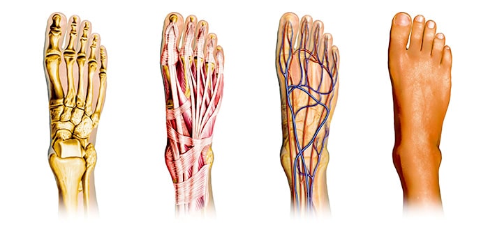
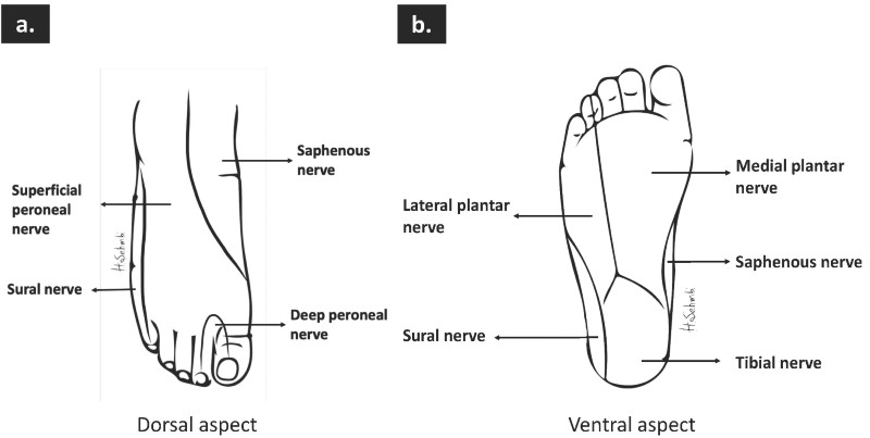
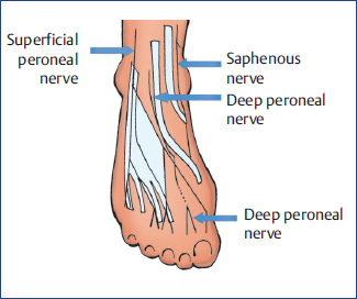
Nerve entrapment at the foot and ankle involves thin and complex anatomic structures and is underdiagnosed because clinical symptoms and electrophysiologic findings may not contribute to the diagno... Upon arising, the sural nerve descends between the heads of the gastrocnemius muscle. It then passes near the lateral margin of the calcaneal tendon, ( running alongside the small saphenous vein until it reaches the ankle. The sural nerve then passes between the lateral malleolus and the calcaneus and enters into the foot. Jan 22, 2022 · Several major nerves serve the human foot. On the top surface of the foot are the dorsal digital nerves and their branches: the deep peroneal nerve, the medial dorsal cutaneous nerve, the intermediate dorsal cutaneous nerve, and the sural nerve. Also on the front surface is the saphenous nerve, which is not included among the dorsal digital nerves because it does not penetrate any of the toes.
Nerves of the foot and ankle diagram. Nerves of the upper limb The upper limb is supplied by a nervous network called the brachial plexus. This plexus is made by the merging of the anterior rami from the lower four cervical nerves and the first thoracic nerve (C5-T1). The plexus is anatomically divided into roots, trunks, divisions, cords and finally, the terminal branches. Neuropathy is a broad term that describes a lack of sensorium, movement, or autonomic function and feedback in a particular area. This can be a centralized neuropathy due to paralysis of extremities via distribution of neurotomes, or a peripheralized neuropathy. Most commonly seen in the extremities is a varying degree of peripheral neuropathy. Peripheral neuropathy usually affects only the ... by M De Maeseneer · 2015 · Cited by 70 — The anatomy of the nerves of the foot and ankle is complex, and familiarity with the normal anatomy and course of these nerves as well as common anatomic ... The nerves of the foot assistance move the body and keep balance both while it’s moving and at rest. All these nerves extend as branches of nerves in the leg that travel through the ankle and into the foot. The sural nerve branches from the tibial and common fibular nerves and is responsible for feeling on the outside of the foot and the little toe. The median and lateral plantar nerves are the 2 biggest nerves in the bottom of the foot.
Anatomy. Function. Associated Conditions. Treatment. The femoral nerve is the major nerve in your thigh. It's one of the largest leg nerves and runs from your pelvis down the front of your leg. The nerve signals carried by the femoral nerve are a critical part of the ability to stand, walk, and maintain balance. 1. Download scientific diagram | Anatomical dissection of the cutaneous nerves of the foot and ankle. 1 Superficial peroneal nerve, 2 Fascial piercing of the superficial peroneal nerve, 3 Superficial ... The joints and muscles of the ankle and foot need to be maintained properly. Nerves provide the ankle and foot with sensation and also tell the muscles when to contract and when to relax. The ankle and foot require nerve supply to function properly. Here’s a look at the nerves that keep the ... Foot Pain Diagram. Written By: Chloe Wilson BSc(Hons) Physiotherapy Reviewed By: FPE Medical Review Board A foot pain diagram is a great tool to help you work out what is causing your ankle and foot pain. There are a whole range of structures e.g. bones, muscles, tendons and nerves which will each give slightly different foot pain symptoms.
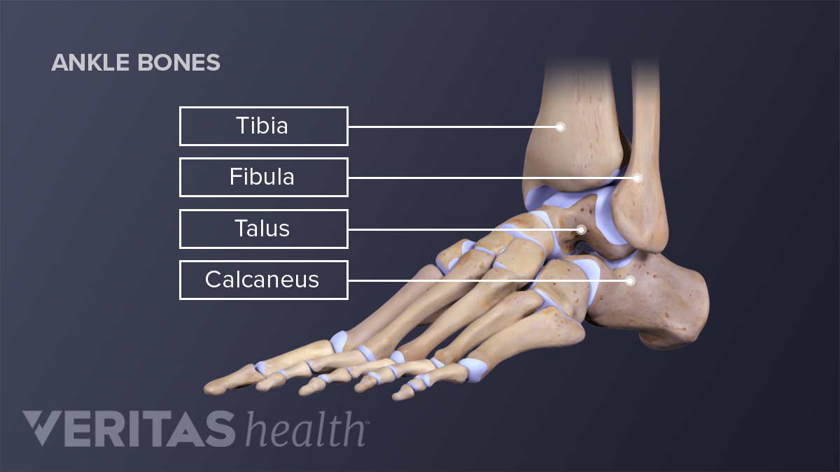
Knee tendons medical vector illustration scheme, anatomical diagram. Tendon, tissue that attaches a muscle to other body parts, usually bones. If you are fortunate, you. Achilles tendon tearing, a common running injury. Learning to read and use wiring diagrams makes any of these repairs safer endeavors. Movahedi Yeganeh M. Triple Tendon Transfer for Correction of Foot Deformity in Common Peroneal Nerve Palsy. Foot Ankle Int. 2016 Jun. 37 (6) ... Diagram of ground reaction vector during heel strike. November 23, 2020 - Calcaneus: the heel bone and the largest bone of the foot. Talus: also called the ankle bone, sits above the heel bone (calcaneus) and makes up the lower part of the ankle joint by connecting the tibia and fibula with the foot. The femoral nerve is located in the pelvis and goes down the front of the leg. It helps the muscles move the hip and straighten the leg. It provides feeling (sensation) to the front of the thigh and part of the lower leg. A nerve is made up of many fibers, called axons, surrounded by insulation, called the myelin sheath.
Foot and Ankle Pain Treatment in Schertz | BioMotion PT
The Danger of Damage During Nerve Entrapment Surgery. If your ankle injury required surgery, it is possible that a nerve was damaged during the procedure. Nerves may also become entrapped in scar tissue following a surgical procedure. In these cases, the surgery intended to correct the problems caused by the ankle sprain has introduced a new ...

The leg, ankle, and foot - Knowledge @ AMBOSS
The nervesof the ankle are derived from the deep and superficial peroneal nerves, the tibial nerves, and the sural and saphenous nerves. The Foot Bones of the foot as seen from the medial (arch) side. The foot is a firm platform that support the weight of the body.

Foot and Ankle Anatomy - Bones, Muscles, Ligaments & Tendons
Dorsal, plantar, medial, and lateral foot perforators are the main groups of PVs in the foot.28 A large PV runs between the first and second metatarsal bones and connects the superficial dorsal venous arch to the pedal vein.29 Clusters of PVs at the ankle are the anterior, medial, and lateral ankle perforators (see Figure 2.13).30 The medial ...
Lower limb arteries and nerves: Anatomy, branches | Kenhub
August 30, 2018 - Main innervation of foot is done by the: Tibial Nerve Deep Fibular Nerve Superficial fibular Nerve Sural Nerve Saphenous Nerves The tibial nerve travels inside the foot via the tarsal tunnel…
Medial Plantar Nerve - Physiopedia
Definition (MEDLINEPLUS) Each of your feet has 26 bones, 33 joints, and more than 100 tendons, muscles, and ligaments. No wonder a lot of things can go wrong. Here are a few common problems: Bunions - hard, painful bumps on the big toe joint. Calluses and corns - thickened skin from friction or pressure.
The Foot – Advanced Anatomy 2nd. Ed.
10 Reviews: Best Foot Creams for Neuropathy (Jan 2022) Say goodbye to the nerve pain that makes your life tough with one of the creams include on our list below. by S. M. Rosyida · updated on Jan 01, 2022 · price $18.78 - $34.95 · 531 views · 32 visitors liked products on this page We hope you love the shops and products we recommend!

14,126 Foot Anatomy Stock Photos, Pictures & Royalty-Free ...
If you still feel apprehensive or are having pain, physical therapy is recommended to retrain the ankle. When a ligament is torn, the nerves that supply the ligament are also injured, contributing to apprehensiveness. Therapy will focus on balance and proprioception (an awareness of where your foot is in space).

Foot and Ankle Anatomy Model
14. Di Er Li Dui. Location: This is another of the pressure points on feet that is located in the toe area. It can be found on the upper side of your second toe, just below your toenail. Uses: Appetite, hiccups and nausea. 15. Di San Li Dui. Location: This point is found right below the toenail of your middle toe.

References in Nerve Disorders of the Hallux - Foot and Ankle ...
Types Of Nerve Injuries - 17 images - ppt nerve injuries powerpoint presentation free, nerve injuries treatment manhattan brooklyn new york, stock illustration of nerve injury type 1 neuropraxia, injuries treated with kinesiology tape are recordable,
Ankle Joint Anatomy and Osteoarthritis
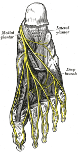
Nerves of the Foot. There are five main nerves that run past the ankle into the foot (Figure 17). All five of these are derived from two nerves that originate from the lumbar spine. The sciatic nerve branches into four of the five primary nerves of the foot. Two segments of the sciatic nerve branch before the knee joint: the tibial nerve and peroneal nerve.

How to Keep Walking With MS Foot Drop | Everyday Health
July 6, 2019 - Nerve pain in foot & peripheral neuropathy cause pain, weakness, pins and needles or numbness. Find out about possible causes, symptoms, diagnosis & treatment.
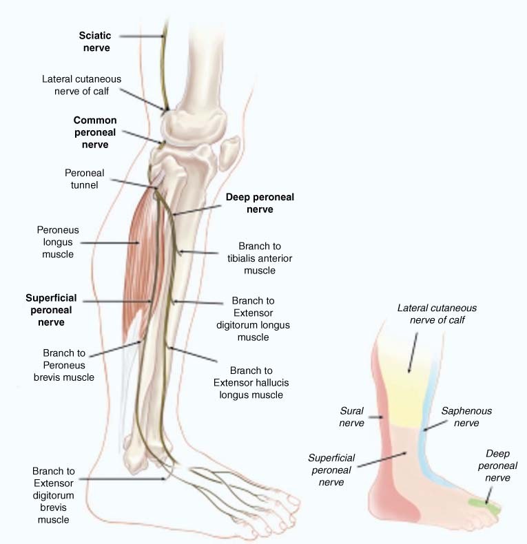
Figure 4 | Nerve Entrapments of the Lower Leg, Ankle and Foot ...
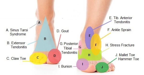
Match the corresponding numbers on the foot diagram below for a list of conditions that may be causing your foot and ankle pain. This is meant for educational purposes only. If you're having a problem with your foot or ankle, visit a podiatrist - a foot and ankle specialist! Top (Dorsal) View of Foot & Ankle Number 1 and 2:

Nerves Of The Leg & Foot - Everything You Need To Know - Dr. Nabil Ebraheim
by M De Maeseneer · 2015 · Cited by 70 — It divides into several thin branches known as dorsal interosseous nerves (1). The lateral branch also provides sensory innervation to the ankle ...

Ankle Block - Landmarks and Nerve Stimulator Technique - NYSORA
Isolated dislocation of the cuboid is a rare injury, and can be easily overlooked at initial presentation. A favourable outcome relies on an accurate and stable reduction. This report documents one such case, successfully treated using an open joystick-assisted reduction and temporary percutaneous Kirschner wire stabilization.
How I Do It: Ultrasound-Guided Ankle Block
Dr. Ebraheim’s educational animated video describes the nerves of the lower leg in a very easy and simple animation.Lateral cutaneous nerve of the calfSural ...

Ankle Block - Landmarks and Nerve Stimulator Technique - NYSORA
A Complete Guide To The Nerves In Your Feet. Problems with nerves in the feet are very common. Many times, an injured nerve will cause intense pain and heat felt within the foot. Nerves act as a network, communicating important information from the foot to the brain. Learn more about the various conditions and problems that can affect the ...

Physical Therapy in Vero Beach for Foot - Anatomy
Advanced Foot And Ankle Treatments In Ann Arbor, MI. Our Board Certified podiatrist in Ann Arbor, MI takes pride in offering the best care and state-of-the-art treatments. Our foot and ankle surgery is done in a professional and comfortable environment. Evaluating and treating your physical condition is part of a treatment plan.

Foot and Ankle | Musculoskeletal Key
Charcot foot is a rare and disabling disorder. It is a result of nerve damage to the feet. A common cause is peripheral neuropathy. Diabetes is the most common cause of this type of nerve damage. This damage is more common in people with type 1 diabetes.
Anatomy of the Foot and Ankle with Foot Drop Deformity Stock ...
"Untreated or repeated ankle sprains may lead to chronic ankle instability, a condition that causes persistent pain and a 'giving way' of the ankle." • A stress fracture may feel like an ankle sprain initially, but you'll also notice swelling without bruising, and pain during normal activities or when touching the area.

Anatomy of the Foot and Ankle | OrthoPaedia
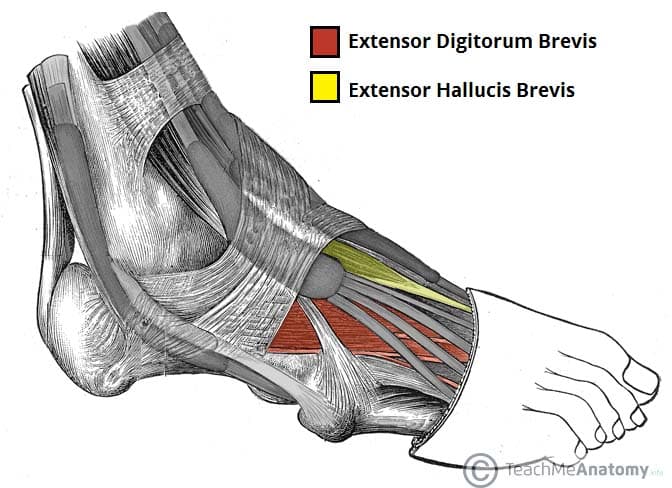
Extensor digitorum brevis, Origin: Superolateral surface of calcaneus bone. Insertion: Middle phalanges of toes 2-4. Innervation: Deep fibular/peroneal nerve ( ...
An MRI study of the tibial nerve in the ankle canal and its ...
Sesamoiditis. Sesamoiditis is an inflammation of the sesamoid bones in the ball of the foot and the tendons they are embedded in. It's usually caused by overuse, especially by dancers, runners and athletes who frequently bear weight on the balls of their feet. It's treated with rest and anti-inflammatory medication.

Nerves and arteries of the foot (preview) - Human Anatomy | Kenhub
October 13, 2017 - Regional anesthesia is increasing in its utility in the treatment of injuries, foreign body removal, and even in the operative setting. By providing a regional block, the affected anatomy can be properly cleaned, explored, and treated without causing unnecessary pain to the patient.

Fysiurgisk Massør | Tag hånd om din krop | Foot anatomy, Body ...
September 20, 2019 - The multidisciplinary foot and ankle team at UC San Diego Health provides the latest diagnostic tests and nonsurgical and surgical therapies to treat minor and complex nerve disorders of the foot and ankle.
In the Foot and Ankle Operation Theater | Musculoskeletal Key
One of the nerves which provides sensation to the bottom of the foot in the tarsal tunnel is the tibial nerve. When this it is compressed, the resulting condition is called tarsal tunnel syndrome, one of the possible reasons for your pinched nerve in foot. Tarsal tunnel syndrome produces symptoms along the nerve that runs from inside the ankle ...
Muscles of the Foot - Dorsal - Plantar - TeachMeAnatomy
Problems with nerves in the foot are very common. Learn about common issues, including causes & treatment, in this complete medical guide to foot nerves.

Physical Therapy in Conway for Ankle Pain - Anatomy
I believe Foot Levelers are actually the number one selling "orthoses" in the country. Some chiros dispense these for everything from headaches to hemorrhoids. My friend is a chiro and is truly embarrassed by Foot Levelers. As far as nerve surgery goes….they are lawsuits and CRPS waiting to happen.

Medial Ankle Pain: Tarsal Tunnel Syndrome | Sports Injury Physio
Shelby Miller Date: January 21, 2022 This diagram shows the parts of the ankle, along with some common problems with the Achilles tendon.. The anatomy of the ankle includes of all structures contained in and surrounding the ankle, or talocrural, joint. These include the contents of the joint capsule such as the ends of the articulating bones, joint cartilage, and synovial fluid.

Muscles, Arteries, and Nerves of Front of Ankle and Dorsum of ...
February 7, 2021 - The foot receives its nerve supply from the superficial peroneal (fibular) nerve, deep fibular nerve, tibial nerve (and its branches), sural nerve, and saphenous nerve. These nerves come from peripheral nerves that arise from the L4 to S3 nerve roots and contribute to the somatic motor function, ...

Ankle Block - Landmarks and Nerve Stimulator Technique - NYSORA
Medial Calcaneal Nerve Entrapment. Medial calcaneal nerve entrapment, often called 'Baxter's nerve' has similar symptoms to that of tarsal tunnel syndrome. A burning pain on the inside of the ankle, below the medial malleolus (bony bit on the inside of the ankle). Pain radiates under the heel and into the arch of the foot.

A Patient's Guide to Foot Anatomy | 2020 OrthoNorCal, Los ...
A serious and potentially life-threatening cause of leg pain is known as a deep vein thrombosis (DVT). This occurs when a clot in a leg vein breaks off and travels to the lungs. Symptoms. In addition to cramping calf pain, other symptoms of a DVT in the lower leg include: Swelling.
Muscles, Arteries, and Nerves of Front of Ankle and Dorsum of ...
Tarsal tunnel syndrome can sometimes result in inner ankle pain as well. This is a condition in which the tibial nerve, which passes behind the medial malleolus of the tibia, becomes entrapped and inflamed, usually resulting in pain and tingling in the foot, although severe impingement of the nerve can be felt as medial ankle pain.
Foot Pain Diagram - Why Does My Foot Hurt?
Menotti F, Laudani L, Damiani A, Orlando P, Macaluso A. Comparison of walking energy cost between an anterior and a posterior ankle-foot orthosis in people with foot drop. J Rehabil Med . 2014 Sep ...

Foot and ankle issues | Northern Arizona Healthcare
August 3, 2018 - The nerves of the foot help move the body and keep balance both while it’s moving and at rest. All of these nerves extend as branches of nerves in the leg that pass through the ankle and into the foot. The sural nerve branches from the tibial and common fibular nerves and is responsible for ...

Anatomical dissection of the cutaneous nerves of the foot and ...
2 weeks ago - The foot contains many bones, muscles, tendons, and other structures. Learn how they work together, plus what can go wrong due to overuse or injury.

Anatomy of the Foot and Ankle (Plantar View) - TrialExhibits Inc.
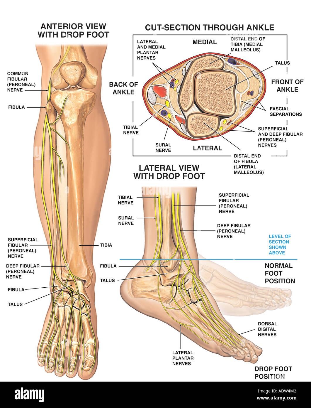
1. Introduction. Foot drop is a general term for difficulty in controlling the ankle-foot complex during the gait. Foot drop is one of the most common secondary conditions following the traumatic injury of the peroneal nerve and stroke [].Partial or total paralysis of the peroneal nerve affects the toes' ability to clear the floor during the swing phase and impairs stability during the stance
Lower limb arteries and nerves: Anatomy, branches | Kenhub
July 6, 2020 - The main nerve to the foot, the posterior tibial nerve, enters the sole of the foot by running behind the inside bump on the ankle (medial malleolus). This nerve supplies sensation to the toes and sole of the foot and controls the muscles of the sole of the foot.

Normal Anatomy and Compression Areas of Nerves of the Foot ...
Jul 03, 2018 · All of these nerves extend as branches of nerves in the leg that pass through the ankle and into the foot. The sural nerve branches from the tibial and common fibular nerves and is responsible for feeling on the outside of the foot and the small toe. The medial and lateral plantar nerves are the two largest nerves in the bottom of the foot.

Foot nerves Images, Stock Photos & Vectors | Shutterstock
Jan 22, 2022 · Several major nerves serve the human foot. On the top surface of the foot are the dorsal digital nerves and their branches: the deep peroneal nerve, the medial dorsal cutaneous nerve, the intermediate dorsal cutaneous nerve, and the sural nerve. Also on the front surface is the saphenous nerve, which is not included among the dorsal digital nerves because it does not penetrate any of the toes.
:max_bytes(150000):strip_icc()/footpainfinal-01-d507e82b3e844d068c0089cbb7004d76.png)
Foot Anatomy, Physiology, and Common Conditions
Upon arising, the sural nerve descends between the heads of the gastrocnemius muscle. It then passes near the lateral margin of the calcaneal tendon, ( running alongside the small saphenous vein until it reaches the ankle. The sural nerve then passes between the lateral malleolus and the calcaneus and enters into the foot.
Comments
Post a Comment