38 onion cell diagram
Let's try how to make onion cell... slide containing red onion cells being observed using a compound light microscope. a) Identify a process that caused the change in the cells. Solution added b) What can be done to restore the cell in the right diagram to its original shape? Base your answers to question 30 on the diagram, which illustrates a transport pathway of CO in the human body. 30. a) Identify the cellular …
Video explaining how to draw a biological diagram showing cell detail.

Onion cell diagram
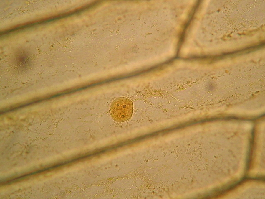
Epidermal Onion Cells Under A Microscope Plant Cells Appear Polygonal From The Cell Diagram Plant Cell Diagram Plant Cell. 62100604d 6wr Fundamental Photographs The Art Of Science Mitosis Mitosis Activity Biology Units. Mitosis In Onion Root Tip Prophase Black Metaphase Green Anaphase Blue Telophase Red Others Are In In Igcse Biology Teaching ... 23.07.2021 · Onion, plain slides, coverslip, watch glass, needles, forceps, brush, blade, safranin, blotting paper, glycerine and compound microscope. THEORY Onion is a multicellular plant. Like other plant cells, the cell of onion peel consists of a cell wall, cell membrane, cytoplasm, a large vacuole and a nucleus. The nucleus lies at the periphery of ... What makes onion cells unique? Onions have a long history of human use, originating in southwestern Asia but having since been cultivated across the world. Their strong odor — actually a defense mechanism — and unique structure belie a complex internal makeup, composed of cell walls, cytoplasm, and the vacuole. What is the difference between […]
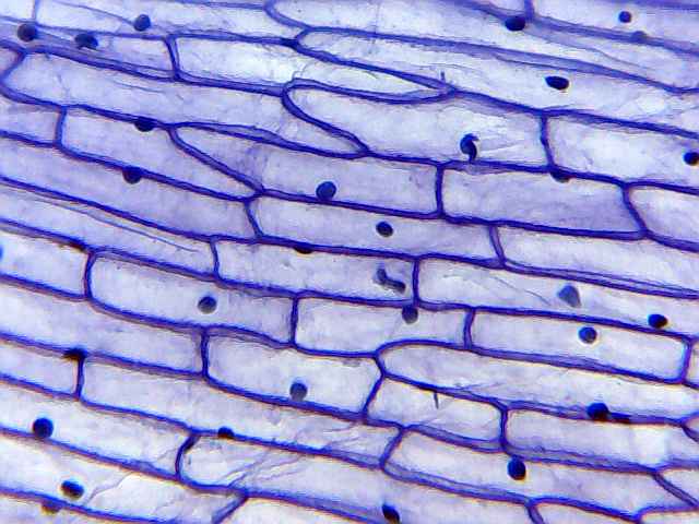
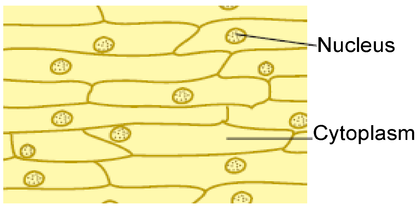
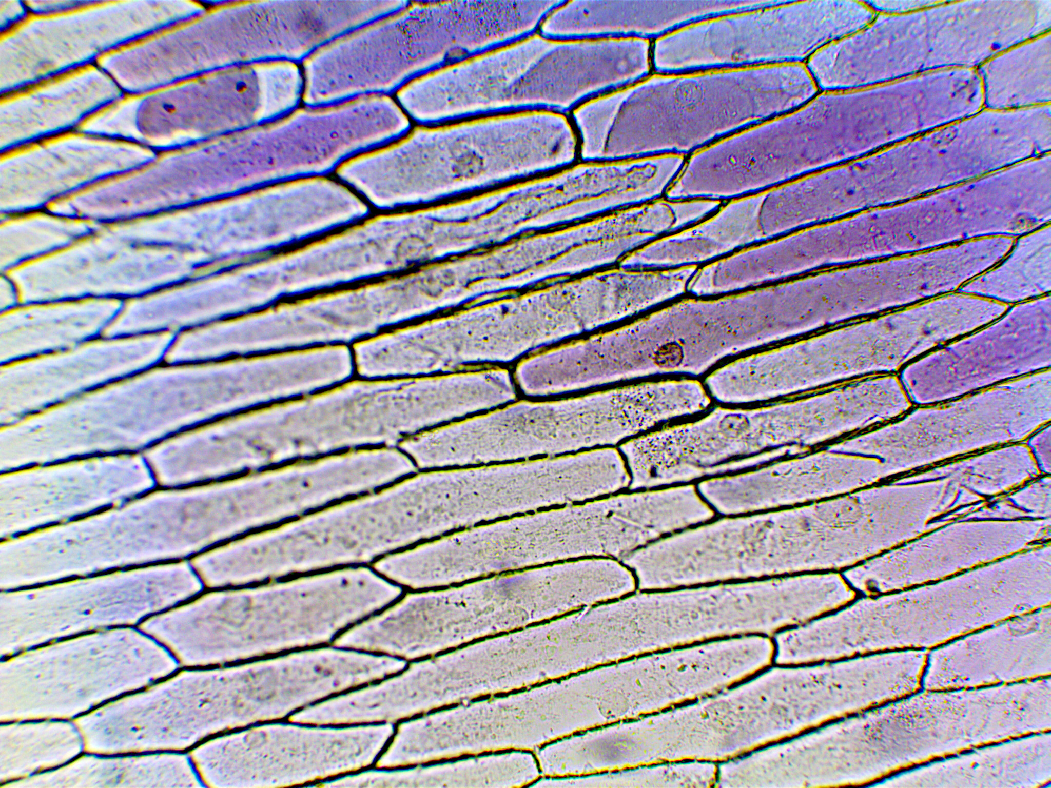
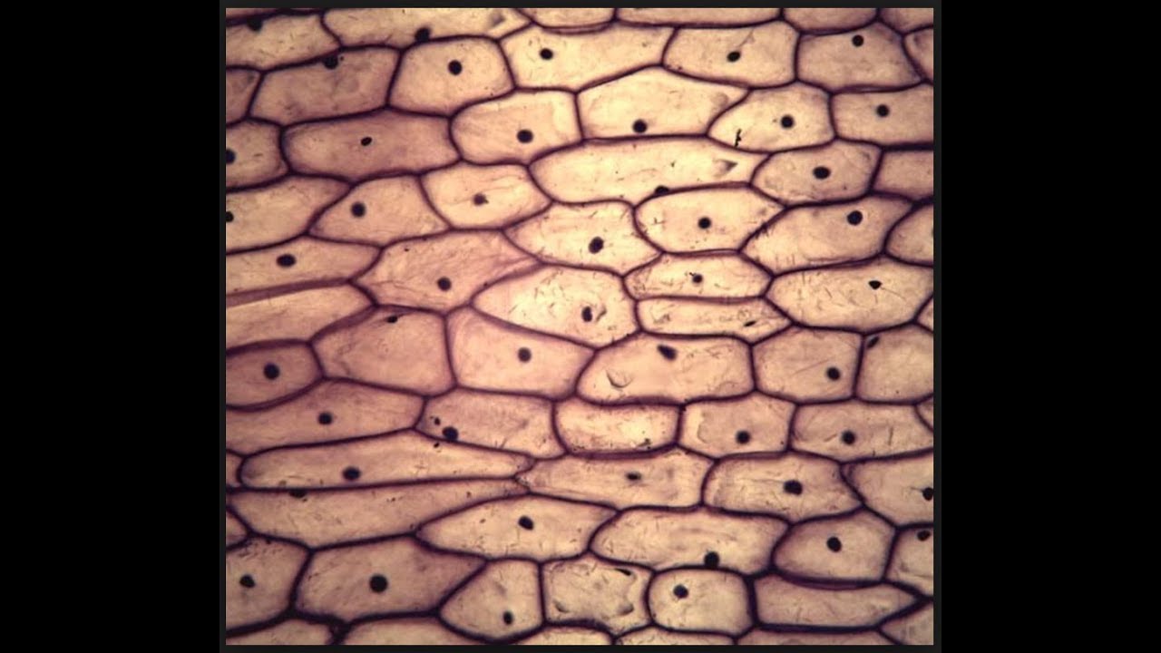
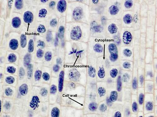
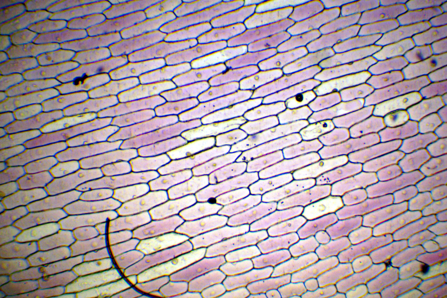
Onion cell diagram. An epidermal onion cell diagram includes components such as citoplasm, a round nucleus and a cell wall. The microscope is the device which helps scientists observe how the onion epidermal layer looks like. Although at the surface an onion has dried protective leaf, inside of it, things change. The onion cell diagram shows that in the middle of. The onion cell diagram shows that in the middle of. The clear epidermal cells exist in a single layer and do not contain chloroplasts, because the onion fruiting body (bulb) is used for storing energy, not photosynthesis. Each plant cell has a cell wall, cell membrane, cytoplasm, nucleus, and a large vacuole. The nucleus is . Prophase: During this first mitotic stage, the nucleolus fades and chromatin (replicated DNA and associated proteins) condenses into chromosomes.Each replicated chromosome comprises two chromatids, both with the same genetic information. Microtubules of the cytoskeleton, responsible for cell shape, motility and attachment to other cells during interphase, disassemble. Staining Onion Cells. Since onion peels are translucent, you’ll need to stain the onion cells before you observe them under the microscope. There are different types of stains depending on what type of cell you are going to look at. Iodine– dark stain that colors starches in cells. In an onion cell, it will make the cell wall more visible.
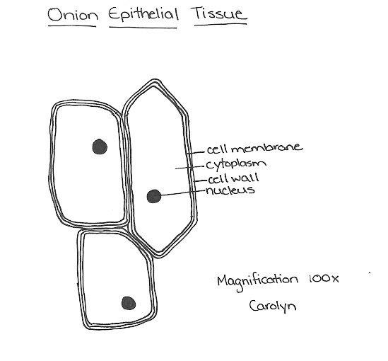
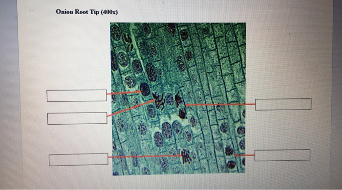
Draw and label the cell wall, cell membrane, cytoplasm, and nucleus. DISCUSSION QUESTIONS. 1. What is the general shape of the onion cells? 2. Describe what you saw without the stain. 3. Why do you think there are many cells close together? 4. Is the onion skin composed of one cell or many cells? 5. Observing onion cells under a microscope is a fun and easy activity for students and hobbyists alike. Onion epidermal cells appear as a single thin layer and look highly organized and structured in terms of shape and size. Certain parts of the cell are also clearly distinguishable with or without staining, making the activity even easier and ... Onion Root Tip Cell Mitosis. Prophase. Nuclear membrane breaks down, chromatin condenses, mitotic spindle forms and attaches to kinetochores. Metaphase. Microtubules align chromosomes along metaphase plate. Anaphase. 22.01.2017 · Cell separation occurs when the individual cell walls are stronger than the adhesive force between them, whereas if the adhesive force is stronger, cell rupture occurs. In general, when the cell walls are rich in pectin, cell wall separation will occur after hydrothermal processing, most likely due to calcium ions in the middle lamella being solubilized by the water …
What makes onion cells unique? Onions have a long history of human use, originating in southwestern Asia but having since been cultivated across the world. Their strong odor — actually a defense mechanism — and unique structure belie a complex internal makeup, composed of cell walls, cytoplasm, and the vacuole. What is the difference between […] 23.07.2021 · Onion, plain slides, coverslip, watch glass, needles, forceps, brush, blade, safranin, blotting paper, glycerine and compound microscope. THEORY Onion is a multicellular plant. Like other plant cells, the cell of onion peel consists of a cell wall, cell membrane, cytoplasm, a large vacuole and a nucleus. The nucleus lies at the periphery of ... Epidermal Onion Cells Under A Microscope Plant Cells Appear Polygonal From The Cell Diagram Plant Cell Diagram Plant Cell. 62100604d 6wr Fundamental Photographs The Art Of Science Mitosis Mitosis Activity Biology Units. Mitosis In Onion Root Tip Prophase Black Metaphase Green Anaphase Blue Telophase Red Others Are In In Igcse Biology Teaching ...
To prepare stained temporary mounts of onion peel - Lab ...

Corona Contact detection app on Android

Onion Peel Cell Diagram With Label - itsessiii

My Drawings from Lab 5 (Cells)
The raw truth about veggies - CSIROscope
9 Reactions of Life Wk1 - Mrs Morritt Science
)
Cellular Structure Of The Onion Skin, Observed Under A ...

The Wonderful Microworld: Cell Nucleus - Onion

ONION SKIN CELLS EPIDERMAL CELLS SHOWS CELL STRUCTURE AND ...

Onion Epidermal Cell Labeled - Top Label Maker

GSEB Solutions for Class 7 Science and Technology - Unit ...
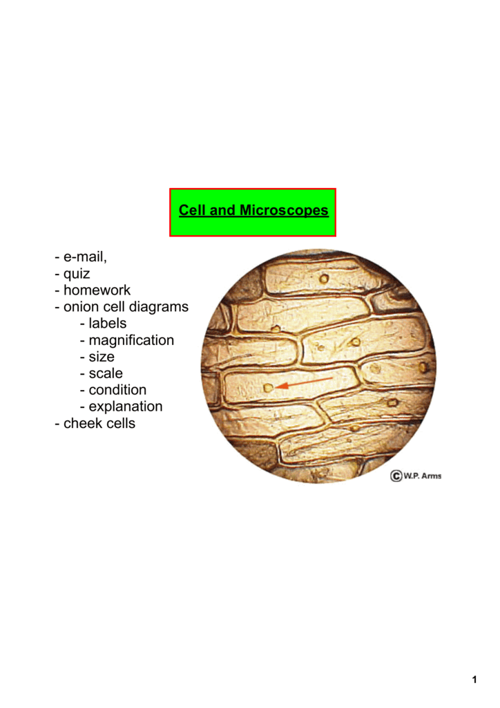
Cell and Microscopes email, quiz homework onion cell diagrams
Microscope Mitochondria In Onion Peel Cell Diagram ...

Pasta Theory À la carte Dark Matter and Energy Being Organic

Image from page 115 of "The Biological bulletin"

Corona contact detection app

Plant Cell Structure Onion Epidermis Photomicrograph 100x ...
![[MN_1993] Onion Cell Diagram Labelled Schematic Wiring](https://static-resources.imageservice.cloud/5235634/cell-microscope-investigation-ppt-video-online-download.jpg)
[MN_1993] Onion Cell Diagram Labelled Schematic Wiring

Biology 109 Lab Review for Practicum #1 - Onion Cells

Image from page 38 of "Advanced biology" (1929)

Blissful Earth: August 2013

Onion Epidermal Cell Labeled - Top Label Maker

Onion Cells | Cell, Biology
Solved: Onion Cell Pictures The Next Two Images Are Real M ...
Onion Epidermal Cell Labeled Diagram
Onion Cells under Microscope

Biology help online: Learning on onion cells

Draw the diagram of cheek cells and label the parts ...
The inner epidermis of the onion bulb's cataphylls (the ...
onion cells - onion cells under microscope - onion cell ...
Lab 11: Mitosis

Iodine Stained Onion Cells - Stock Image - C028/3135 ...

Onion Epidermal Cell Labeled Diagram - Atkinsjewelry
Onion Cells under Microscope

Subcellular localization of wheat ATG8s in onion epidermal ...

Onion Cell Structure Under Microscope - Micropedia

Onion Epidermal Cell Labeled Diagram - Hanenhuusholli

NCERT Class 9 Science Lab Manual - Slide of Onion Peel and ...
Comments
Post a Comment