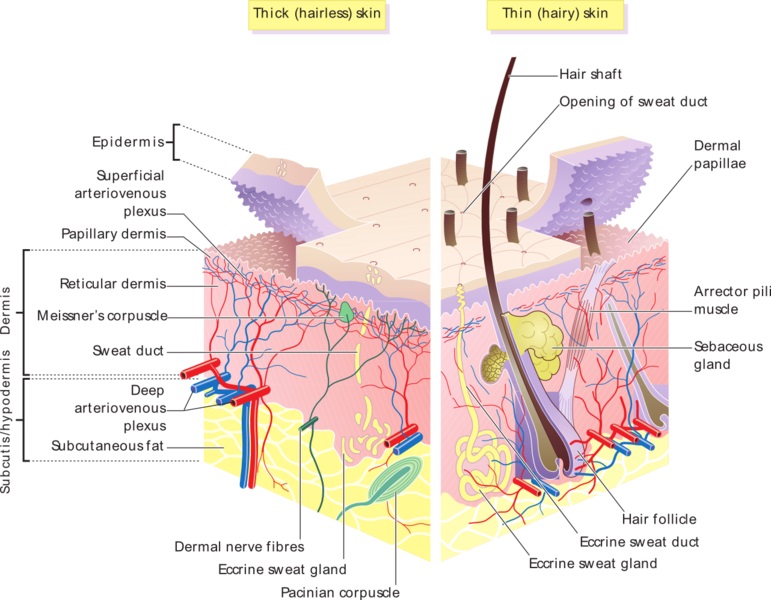
42 diagram of thin skin
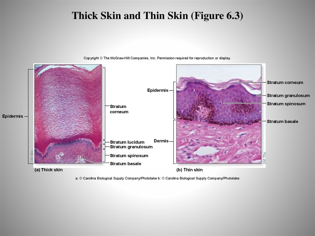
The skin has a surface area of between 16.1–21.5 sq ft. for an adult human. The thickness of the skin differs over all parts of the body, and between men and women and the young and the old. For example, the skin on the forearm which is on average 1.3 mm in the human male and 1.26 mm in … Thin skin has many structures present that are absent in thick skin. Thick skin does have an extra epidermal layer called the stratum lucidum, which is absent in thin skin. The function of thick skin is mainly to prevent damage due to abrasion and friction. The thin skin also functions in protection, but also produces hairs, sweat, and sebum.
Nov 16, 2018 · Diagram Of Thin Skin Structure. 5 1 Layers Of The Skin Anatomy And Physiology. Thick And Thin Skin Structure 14 Download Scientific Diagram. Solved Label The Skin Structures And Areas Indicated In The Ac. Skin Integumentary System.

Diagram of thin skin
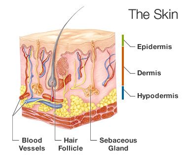
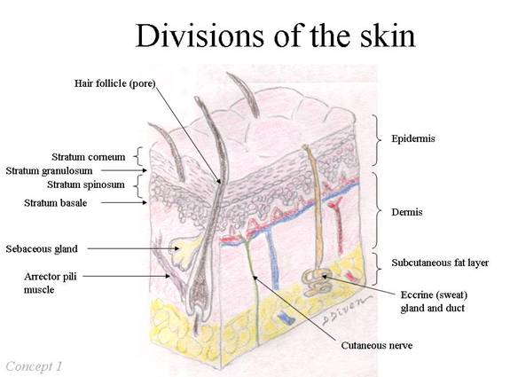
3-D Skin Model Project - Anatomy and Physiology. This is a good basic project to have the students build a 3-D model of the skin. The project encourages students to build a 3-D Skin model to build, and describe the functions of various skin components. The assignment is out of 30 marks and a sample is attached on the second page. The skin is the largest organ of the body, with a total area of about 20 square feet. The skin protects us from microbes and the elements, helps regulate body temperature, and permits the ... Skin is the largest organ in the body and covers the body's entire external surface. It is made up of three layers, the epidermis, dermis, and the hypodermis, all three of which vary significantly in their anatomy and function. The skin's structure is made up of an intricate network which serves as the body's initial barrier against pathogens, UV light, and chemicals, and mechanical injury.
Diagram of thin skin. Label the Skin Structures and areas Indicated In the Accompanying Diagram Of Thin Skin. name lab time date review sheet the integumentary system 4 label the skin structures and areas indicated in the ac panying diagram of thin skin then plete the statements that follow a granules extruded wbdg the gateway to up to date information on integrated whole building design techniques and technologies ... Anatomy of the Skin. The skin is a vital organ that covers the entire outside of the body, forming a protective barrier against pathogens and injuries from the environment. The skin is the body's largest organ; covering the entire outside of the body, it is about 2 mm thick and weighs approximately six pounds. Label the skin structures and areas indicated in the accompanying diagram of thin skin. Label the skin structures and areas indicated in the accompanying diagram of thin skin. Then complete the statements that follow hair shaft stratum corneum granulosum stratum stratum spinosum stratum basale layers papillary layer denna. Terms in this set 64 ... The dermis is the middle layer of the three layers of skin. It's located between the epidermis and the subcutaneous tissue. It contains connective tissue, blood capillaries, oil and sweat glands, nerve endings, and hair follicles. The dermis is split into two parts—the papillary dermis, which is the thin, upper layer, and the reticular dermis ...
Anatomy & Physiology continues with a look at your biggest organ - your skin.Pssst... we made flashcards to help you review the content in this episode! Find... Skin Definition. Skin is the soft outer tissue which covers vertebrates. In humans, it is the body's largest organ, covering a total area of about 20 square feet.It protects our internal organs from the environment using a multi-layered system of cushioning, a cellular barrier, and protective oils. Sebaceous Glands. A sebaceous gland is a type of oil gland that is found all over the body and helps to lubricate and waterproof the skin and hair. Most sebaceous glands are associated with hair follicles. They generate and excrete sebum, a mixture of lipids, onto the skin surface, thereby naturally lubricating the dry and dead layer of keratinized cells of the stratum corneum, keeping it pliable. Children have thin skin, which gradually thickens until the fourth decade of life, affected by the concentration of sex steroids, general health, and hydration. The skin begins to thin again during the fifth decade of life, primarily due to changes in the dermis with loss of epithelial appendages, elastic fibers, and ground substance, among others.
In the diagram of skin shown below, which structure illustrates that thin skin is present? a) C b) D c) E d) F e) H. a) a. In the diagram of skin shown above, which labeled structure generates fingerprints? a) A b) B c) G d) D. c) g. In the diagram of skin shown above, which area is primarily composed of areolar connective tissue? a) E b) F c) G Skin that has four layers of cells is referred to as "thin skin." From deep to superficial, these layers are the stratum basale, stratum spinosum, stratum granulosum, and stratum corneum. Most of the skin can be classified as thin skin. "Thick skin" is found only on the palms of the hands and the soles of the feet. Skin (Integumentary System) Blogger: Scientist. Thin Skin (Hematoxylin-Eosin) Thin Skin (Mallory Trichrome) Thin Skin (Verhoeff-Van Gieson) Differences between thick and thin skin in light microscope specimens. Share. CHAPTER 1. ANATOMY AND PHYSIOLOGY OF THE SKIN 3 or stratum spinosum (Murphy, 1997). The squamous layer is composed of a variety of cells that differ in shape, structure, and subcellular properties depending on their location. Supra - basal spinous cells, for example, are polyhedral in shape and have a rounded nucleus, whereas cells of the upper ...
5 1 Layers Of The Skin Anatomy Physiology
30.4.2020 · Dermatomes are areas of skin, and each communicates with the brain via a single nerve. Here, find out more about the relationship between nerves and dermatomes.

Amazon Com Skin Care Chart 2 Sided Laminated Quick Reference Guide Covers Skin Care Services From Skin Analysis Facials Waxing Eye Brow Shaping Massage Anatomy Industrial Scientific
Label the skin structures and areas indicated in the accompanying diagram of thin skin. Then, complete the statements that follow. a. Lamellar granules contain glycolipids that prevent water loss from the skin. b. Fibers in the dermis are produced by fibroblasts .
Skin Diagram Hair Shaft Stratum Corneum Epidermis Stratum Basale Ppt Video Online Download
In the thick skin on the soles of the feet and palms of the hands the stratum lucidum is a thin, transparent layer of dead keratinocytes lying superficial to the stratum granulosum. The outermost layer is the stratum corneum, a thick layer of dead, flattened, keratin-filled keratinocytes that protect the underlying tissues.

Jaypeedigital Ebook Reader
In the diagram of skin shown below, which structure illustrates that thin skin is present? C. In the diagram of skin shown below, which area is primarily composed of areolar connective tissue? G. In the diagram of skin shown below, where is smooth muscle found? D.
Tissue Models For Rf Exposure Evaluation At Frequencies Above 6 Ghz Ziskin 2018 Bioelectromagnetics Wiley Online Library
15.3.2017 · Trachea Anatomy and Structure Tracheal Tissues and Membranes. Respiratory Mucosa: The innermost layer of the trachea, consisting of ciliated pseudostratified columnar epithelium and lamina propria (a thin layer of connective tissue), is covered with a sticky mucus coating produced by the goblet cells present in the region [1].
Hand Skin Grafts Florida Orthopaedic Institute
The skin is considered thick or thin depending on the thickness of the epidermis. It has a complex structure made of networks of cells, muscles, and nerves to protect the body from pathogens and environmental damage. The different thicknesses across the body parts to maintain different body functions and keep you healthy.
What Is The Difference Between Thick And Thin Skin Pediaa Com
Thin skin is a common condition in older adults, and is most noticeable in the face, arms, and hands. Treatment can prevent thin skin from getting worse.

Skin Integumentary System
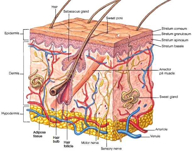
Skin that has four layers of cells is referred to as “thin skin.”. From deep to superficial, these layers are the stratum basale, stratum spinosum, stratum granulosum, and stratum corneum. Most of the skin can be classified as thin skin. “Thick skin” is found only on the palms of the hands and the soles of the feet.

Jaypeedigital Ebook Reader
Skin can either be thin or thick. The main difference is the thickness of the epidermis and dermis, which are the top two layers of skin. Thin skin covers most of the body and can vary in thinness ...

Human Anatomy Physiology 1
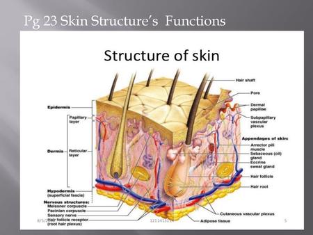
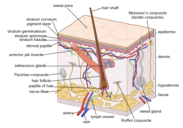
Three layers of skin: The epidermis: a thin outer portion, that is the keratinised stratified squamous epithelium of skin. The epidermis is important for the protective function of skin. The basal layers of this epithelium are folded to form dermal papillae. Thin skin contains four types of cellular layers, and thick skin contains five.

All You Need To Know About Thin Skin Treatment Causes Symptoms Prevention Healthcare Business Today
7.9.2021 · Ciliated epithelium is an important tissue found in various parts of the body and aids in everyday health. Explore what ciliated epithelial is and its function, its structure using a diagram, why ...

Photomicrographs Of Thin Skin Sections A A Section In Thin Skin Of Download Scientific Diagram
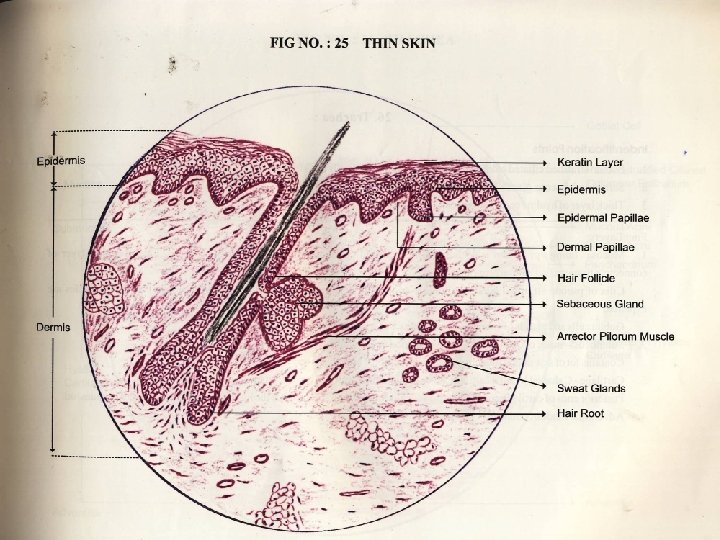
Structure of thin skin of an animal. In the structure of the thin skin of animals, you will find the two distinct layers - the epidermis and dermis. I will show you the different histological characteristics from the epidermis and dermis of a skin microscope slide. #1. The epidermis of skin (you might learn the general features, layers of the ...

Integument Thin Skin Model Skin Model Skin Anatomy Skin
In deserts, the human skin gets thicker to prevent water loss to dry air. Organisms with thin skin have the possibility of losing water all the time and need to stay near water to prevent it from drying. Sensation. Skin is the main sense organ that can sense touch, heat, pressure, cold, pain, and pleasure.

Somatosensory Receptors Biology For Majors Ii
referred to as thin skin. In areas of the body exposed to greater friction, like the fingertips, palms and soles of the feet the epidermis has five strata or layers. This is referred to as thick skin.
Human Skin Wikipedia
Eye Diagram. The eye – one of the most complex organisms in the human body. It is made up of many different parts working in unison together. In order for the eye to work at its best, ... An eyelid is a thin fold of skin that covers and protects the eye. Pupil.

Thin Skin Layers Diagram Quizlet
skin •Paper-thin skin •Dark or reddened areas 18 Darkly pigmented skin does not blanch. Parameter 3: Skin Color Redness •Reddened skin on the sacral area can be from a variety of etiologies. ... -Diagram of a body outline where staff can note any skin changes they observe. 30.

Thin Skin Versus Thick Skin Histology Skin Anatomy Thick Skin Integumentary System
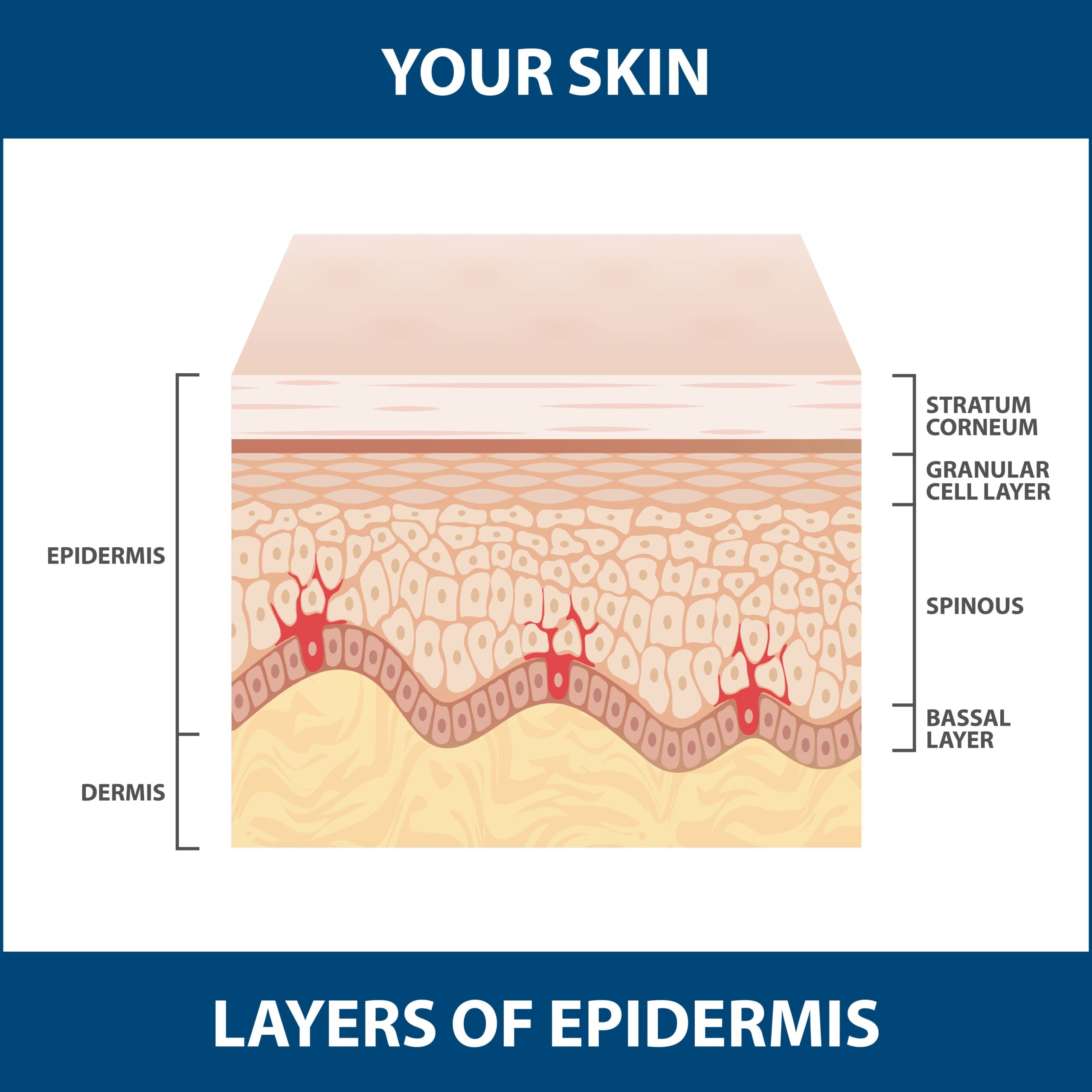
This diagram shows schematically, the four different layers found in the epidermis of most skin (thin skin). This epidermis of skin is a keratinized, stratified, squamous epithelium . Cells divide in the basal layer, and move up through the layers above, changing their appearance as …
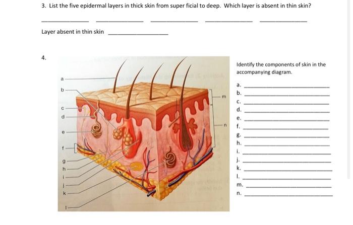
Solved 3 List The Five Epidermal Layers In Thick Skin From Chegg Com
30.7.2020 · The heart is a muscular organ about the size of a closed fist that functions as the body’s circulatory pump. It takes in deoxygenated blood through the veins and delivers it to the lungs for oxygenation before pumping it into the various arteries (which provide oxygen and nutrients to body tissues by transporting the blood throughout the body).
Skin Information Layers Of Skin Keeping Skin Healthy And More
6.12.2017 · The thin superior part that blends with the forehead is called the root of the nose, ... Nose Anatomy Diagram. Does Your Nose Keep Growing All Your Life. ... The trigeminal nerve innervates the outer skin of the human nose [8]. Function of the Nose
Skin The Histology Guide
Download scientific diagram | Thin skin. Human. Light micrograph of a longitudinal section of female cheek showing the four layers of the thin skin and the four different cell types of epidermis.
1
Dermis: Thick skin has a thinner dermis than thin skin, and does not contain hairs, sebaceous glands, or apocrine sweat glands. Thick skin is only found in areas where there is a lot of abrasion - fingertips, palms and the soles of your feet. show labels. This is a picture of an H&E stained section of the epidermis of thin skin.

Layers Of Skin How Many Diagram Model Anatomy In Order
Skin is the largest organ in the body and covers the body's entire external surface. It is made up of three layers, the epidermis, dermis, and the hypodermis, all three of which vary significantly in their anatomy and function. The skin's structure is made up of an intricate network which serves as the body's initial barrier against pathogens, UV light, and chemicals, and mechanical injury.

Layers Of The Skin Anatomy And Physiology I
The skin is the largest organ of the body, with a total area of about 20 square feet. The skin protects us from microbes and the elements, helps regulate body temperature, and permits the ...
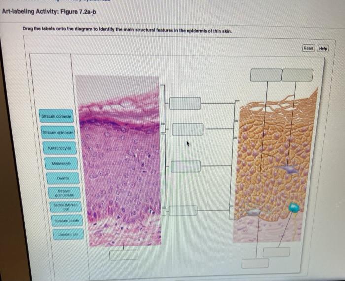
Solved Art Labeling Activity Figure 7 2a B Drag The Labels Chegg Com
3-D Skin Model Project - Anatomy and Physiology. This is a good basic project to have the students build a 3-D model of the skin. The project encourages students to build a 3-D Skin model to build, and describe the functions of various skin components. The assignment is out of 30 marks and a sample is attached on the second page.
Anatomy Of The Skin

Structure And Function Of The Skin Skin Disorders Msd Manual Consumer Version

Calluses On Thin Skin With No Lucidum Google Search Integumentary System Thin Skin Thick Skin
The Integumentary System Online Presentation

5 1 Layers Of The Skin Anatomy Physiology

18 Usmle Dermato Ideas Epidermis Dermis Integumentary System

Dermis Wikipedia
Skin Basic Facts About Skin Largest Organ Of
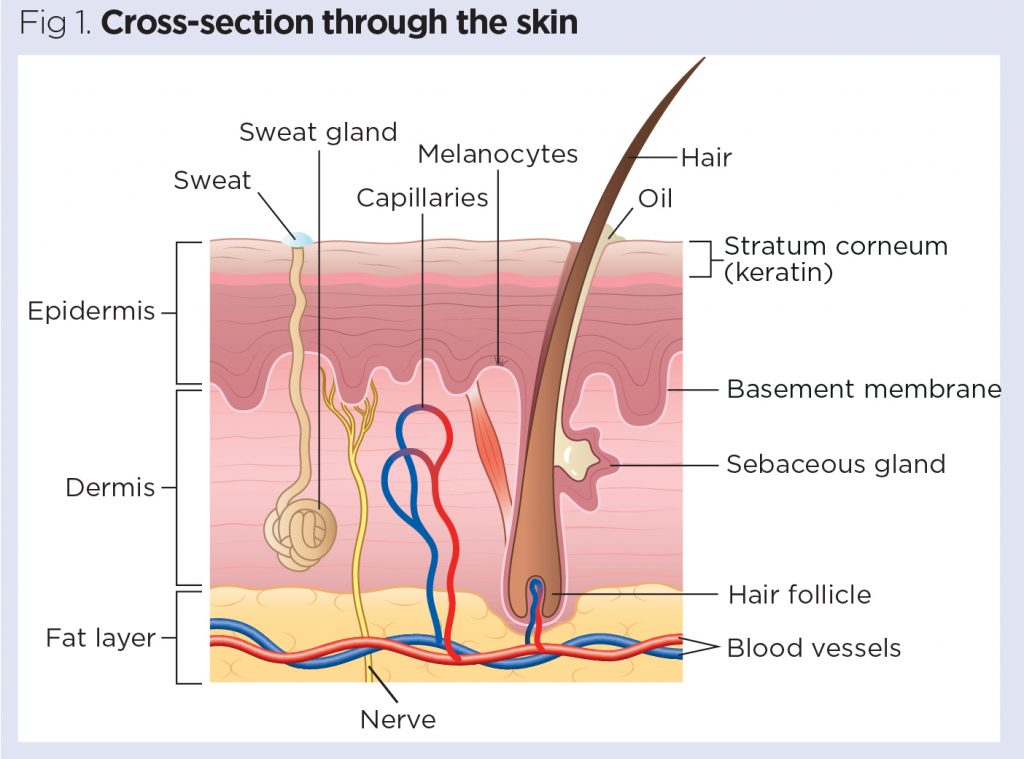
Skin 1 The Structure And Functions Of The Skin Nursing Times
The Integumentary System Skin Medical Terminology For Cancer
1
1

Are Retinol And Hydroxy Acids Thinning Our Skin Eudelo
Skin Cells Layers And Histological Features Kenhub

Layers Of The Skin Anatomy And Physiology I

Thick And Thin Skin Structure 14 Download Scientific Diagram
Skin Cells Layers And Histological Features Kenhub

Histology Of Thin Skin Hairy Skin Youtube
Comments
Post a Comment