41 diagram of amoeba
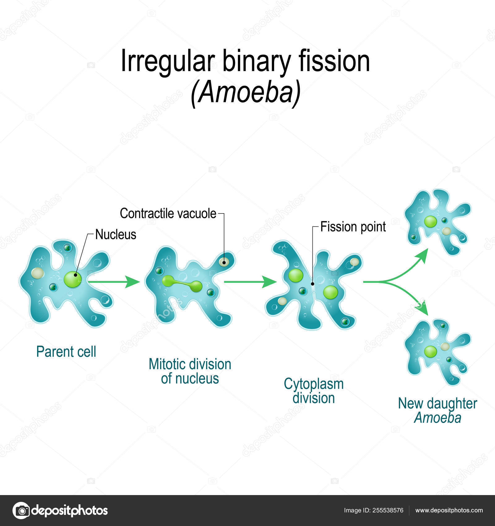
In amoeba, the nucleus in the cell elongates and divide into two parts from the centre. One may also ask, what is binary fission explain with diagram? With the help of suitable diagrams describe binary fission. Binary fission is a kind of asexual reproduction. It is a common type of reproduction found in bacteria and protists like Amoeba in ... Amoeba Under The Microscope Fixing, Staining Techniques and Structure. Amoeba (plural amoebas/amoebae) is a genus that belongs to Kingdom protozoa. Generally, the term is used to describe single celled organisms that move in a primitive crawling manner (by using temporary "false feet" known as pseudopods).
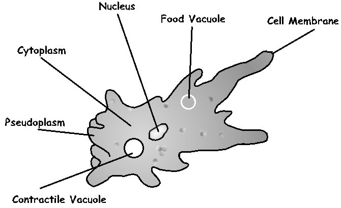
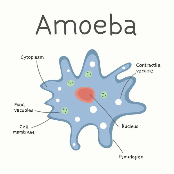
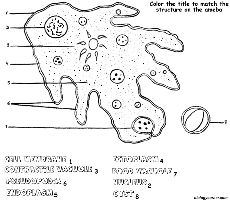
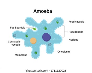
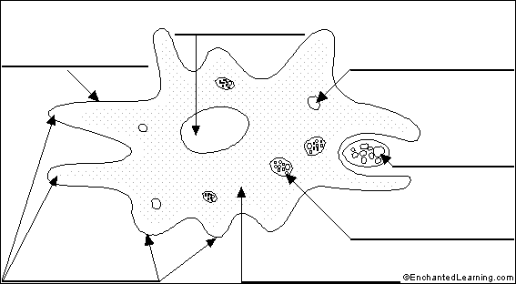
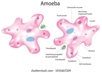
Structure of amoeba primarily encompasses 3 parts - the cytoplasm, plasma membrane and the nucleus. The cytoplasm can be differentiated into 2 layers - the outer ectoplasm and the inner endoplasm. The plasma membrane is a very thin, double-layered membrane composed of protein and lipid molecules.

Diagram of amoeba
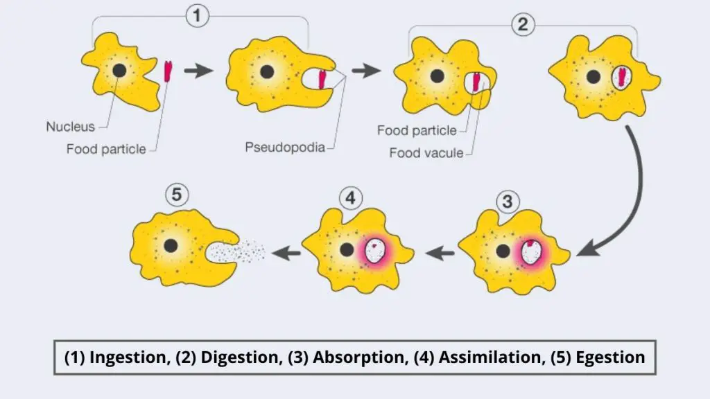
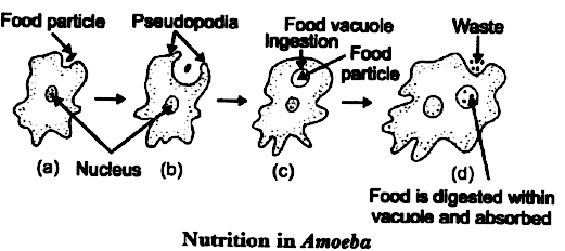
Nutrition in an Amoeba occurs through a process called phagocytosis where the entire organism pretty much engulfs the food it plans on eating up. The mode of nutrition in amoeba is known as holozoic nutrition. It involves the ingestion, digestion and egestion of food material. Amoeba does not have any specialized organ for nutrition. Label Amoeba Anatomy Diagram Printout. EnchantedLearning.com is a user-supported site. As a bonus, site members have access to a banner-ad-free version of the site, with print-friendly pages. The structure and diagram of amoeba. Amoeba. Paramecium Diagram. Body structure of an amoeba proteus. Amoeba. Amoeba. Irregular binary fission. Paramecium Dividing bacteria diagram. Amoeba is a single celled organism that appears transparent. Nutrition in an Amoeba occurs through a process called phagocytosis.
Diagram of amoeba. The amoeba is a tiny, one-celled organism with pseudopods - Kingdom Protista. Label Amoeba. Label Amoeba Anatomy Diagram Printout. Animal Cell Anatomy. Explore autosomal recessive trait and X-linked recessive trait tracking in pedigrees with the Amoeba Sisters! Matching handout available here: http://www.amo... A paramecium is a unicellular (one cell) eukaryotic organism generally found in stagnant water. While very small, sometimes large paramecium can be seen as tiny specks darting around in a water sample. Paramecium can be about 0.5 mm long. Species of Paramecium range in size from 50 to 330 micrometres (0.0020 to 0.0130 in) in length. Genus: Amoeba . Species: proteus. Amoeba proteus is a unicellular organism widely distributed in ponds, lakes, freshwater pools and slow streams. Normally it is found creeping, feeding upon algae, bacteria etc. Under the microscope, it appears as irregular, jelly-like tiny mass of hyaline protoplasm.
The mode of nutrition in amoeba is holozoic. The process of obtaining food is called phagocytosis. Amoeba feeds on microscopic organisms floating on water. Amoeba (Ameba) Animal Printouts. Label Me! Printouts. The amoeba is a tiny, one-celled organism. You need a microscope to see most amoebas - the largest are only about 1 mm across. Amoebas live in fresh water (like puddles and ponds ), in salt water, in wet soil, and in animals (including people). There are many different types of amoebas. An amoeba (/ ə ˈ m iː b ə /; less commonly spelled ameba or amœba; plural am(o)ebas or am(o)ebae / ə ˈ m iː b i /), often called an amoeboid, is a type of cell or unicellular organism which has the ability to alter its shape, primarily by extending and retracting pseudopods. Amoebae do not form a single taxonomic group; instead, they are found in every major lineage of eukaryotic ... Diagram of an Amoeba Moving: Use the internet to research how an amoeba moves. Draw a schematic diagram to show cell movement or how a pseudopod extends outward. Label the diagram. NOTE: There are different ways to get five marks. This question is about researching the process and presenting the findings in a diagram.
An amoeba is an aquatic, single-celled protist characterized by a gelatinous body, amorphous shape, and amoeboid movement. Amoebas can form temporary extensions of their cytoplasm known as pseudopodia or "false feet" which can be used for locomotion or capturing food. Food acquisition is amoebas occurs by a type of endocytosis called phagocytosis. Amoeba is an aquatic, single-cell (unicellular) organism with membrane-bound (eukaryotic) organelles that has no definite shape. It is capable of movement. When seen under a microscope, the cell looks like a tiny blob of colorless jelly with a dark speck inside it. Some parasitic amoebae living inside animal bodies, including humans, can cause various intestinal disorders such as diarrhea ... the direction of flow in passive transport. Show this in the diagram on right by drawing in 10 total circles (to represent molecules). You must decide a certain amount to place on the left vs. the right side after viewing the arrow indicating the direction of movement. Label the high concentration side and low concentration side. Amoeba Diagram to basically explain you about the general definition of amoeba and to provide you with some explanation examples in diagrams. Do you know about thing called amoeba? Ever heard anything about it? In biology learned at the basic school people usually know about this entity called amoeb...
Diagram Of The Dictyostelium Life Cycle Individual Amoebas Feed On Download Scientific Diagram
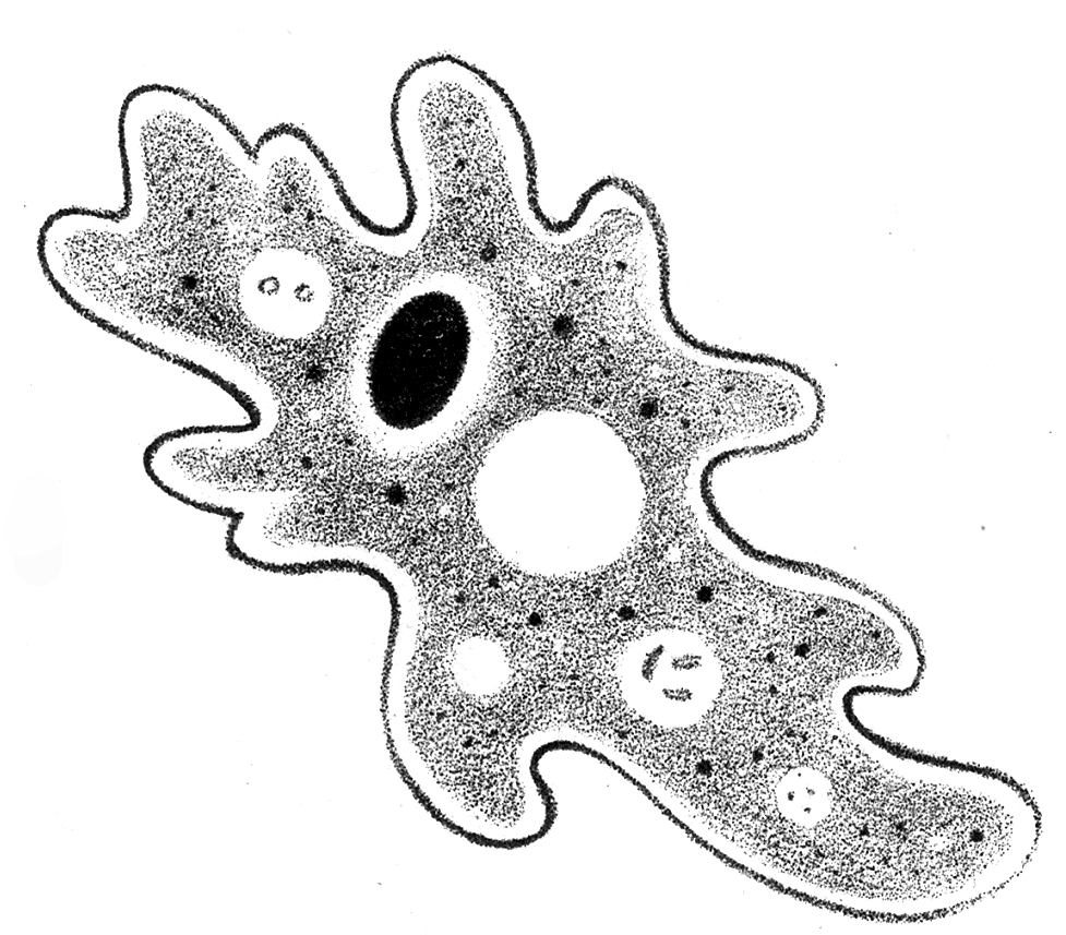
Amoeba proteus is a one-celled animal about 25 mm ( 1/100 inch) in diameter, and is, therefore, invisible to the naked eye. Under the compound microscope it appears as an irregular colorless particle of animated jelly which is constantly changing its shape by thrusting out finger-like processes. Habitat of Amoeba proteus.

How To Draw Amoeba Labelled Science Diagram Easy Way Drawing Amoeba Amoeba Drawing And Parts Lables Youtube Science Diagrams Draw Diagram Diagram
Explore the steps of DNA replication, the enzymes involved, and the difference between the leading and lagging strand! This video is an update from our old D...

Amoeba Diagram Diagram Quizlet
66 Royalty-free Vector Images & Drawings of Amoeba diagram. yaroslav-reis Amoeba - the structure of the microorganism. Vector graphics. Marinka Abstract oval shape tiny protist amoeba organelle pellicle parasite element. Line black hand drawn lab microbe icon sign symbol pictogram diagram sketch Art doodle cartoon style design.
Labeled Amoeba Diagram By Sciencedoodles On Deviantart
Start studying Amoeba Sisters Video Recap: Cell Transport. Learn vocabulary, terms, and more with flashcards, games, and other study tools.
66 Amoeba Diagram Vector Images Amoeba Diagram Illustrations Depositphotos
Download a free printable outline of this video and draw along with us: https://artforall.me/video/how-to-draw-amoeba Thank you for watching. Please subscri...

How Does An Amoeba Obtain Its Lido
How TO Draw amoeba/diagram of amoeba/amoeba drawing, drawing amoeba, drawing of amoeba, easy way of drawing amoeba, protozoa, one cell animal, how to draw di...
35 Diagram Of Amoeba With Label Labels Database 2020
The structure and diagram of amoeba. Vector. Amoeba icon vector from virus collection. Thin line amoeba outline icon vector illustration. Linear symbol for use on web and mobile apps, logo, print media. Balamuthia mandrillaris amoeba. 3D illustration. A free-living protozoan in soil and water, can cause granulomatous amoebic encephalitis of the ...

The Structure And Diagram Of Amoeba Stock Vector Adobe Stock
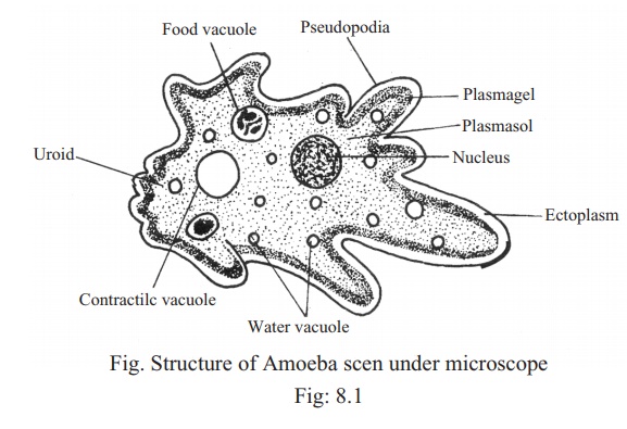
A labelled diagram of Amoeba proteus can be seen above. The pseudopodia are the most defined structures of A. proteus and part of what makes the organism so fascinating. These "false feet" are used for movement and to engulf prey (see Nutrition for further detail) - making it an essential part of its structure.

Amoeba Cell Diagram
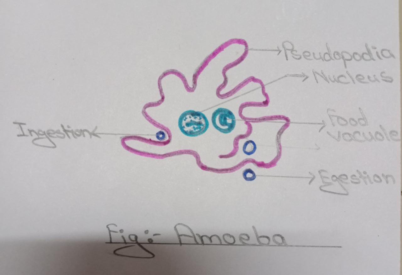
Ingestion. Amoeba has holozoic mode of nutrition. Amoeba is a unicellular organism and thus has no specialised organs or structures for the process of nutrition. It takes place through the general body surface through pseudopodia. Amoeba intakes the food by the process of invagination.

Amoeba Genus Classification Structure All Life Processes
ADVERTISEMENTS: In this article we will discuss about the structure of amoeba. This will also help you to draw the structure and diagram of amoeba. 1. Fresh water and free living organism commonly available in stagnant water. ADVERTISEMENTS: 2. Body irregular and cytoplasm clearly differentiated into ectoplasm and endoplasm. 3. Body naked, and extends into numerous […]
Amoeba Structure Images Stock Photos Vectors Shutterstock
A labelled diagram of Amoeba proteus can be seen above. Pseudopodia (written as pseudopod on the picture) are temporary finger like projections with blunt rounded tips which are constantly being given out or withdrawn by the body. Many pseudopodia are formed simultaneously. Amoeba exhibits movement by the pseudopodia. It also helps in food capture.

Draw The Labelled Diagram Amoeba
An amoeba is unicellular and moves by using pseudopods. A pseudopod is a temporary bulge that forms in the cell membrane as a result of the movement of the cytoplasm. The word pseduopod means "false foot." The pseudopod has two functions, or uses: 1. to move, 2. to capture food.
Label Amoeba Enchantedlearning Com
Amoeba cell diagram. An amoeba is a microscopic organism with a wacky, constantly-changing shape. It can be dangerous once it infects a human host, although by observing proper safety guidelines at your lab, it'll be perfectly fine to observe and experiment on these microbes. Amoebae are a type of eukaryotic organism made up of only a single ...
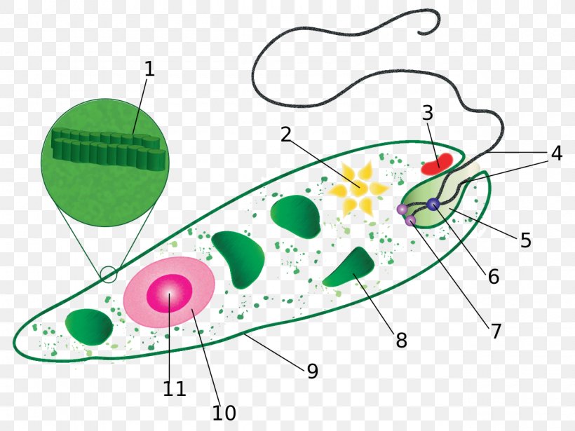
Euglena Diagram Protist Flagellate Cell Png 1280x960px Euglena Amoeba Anatomy Area Asexual Reproduction Download Free
Amoeba. Body structure: Amoeba appears as a colourless and transparent drop of jellywhen viewed under a microscope. It lacks a definite body shape because it changes its shape by producing the pseudopodia every moment. So, it is not possible to describe its definite shape, anterior or posterior ends, dorsal and ventral surfaces.

How To Draw Amoeba Labeled Science Diagram Youtube
Search from Diagram Of An Amoeba Pic stock photos, pictures and royalty-free images from iStock. Find high-quality stock photos that you won't find anywhere else.
Amoeba Cell Characteristics Structure Movement Nutrition Reproduction Disease Habitat
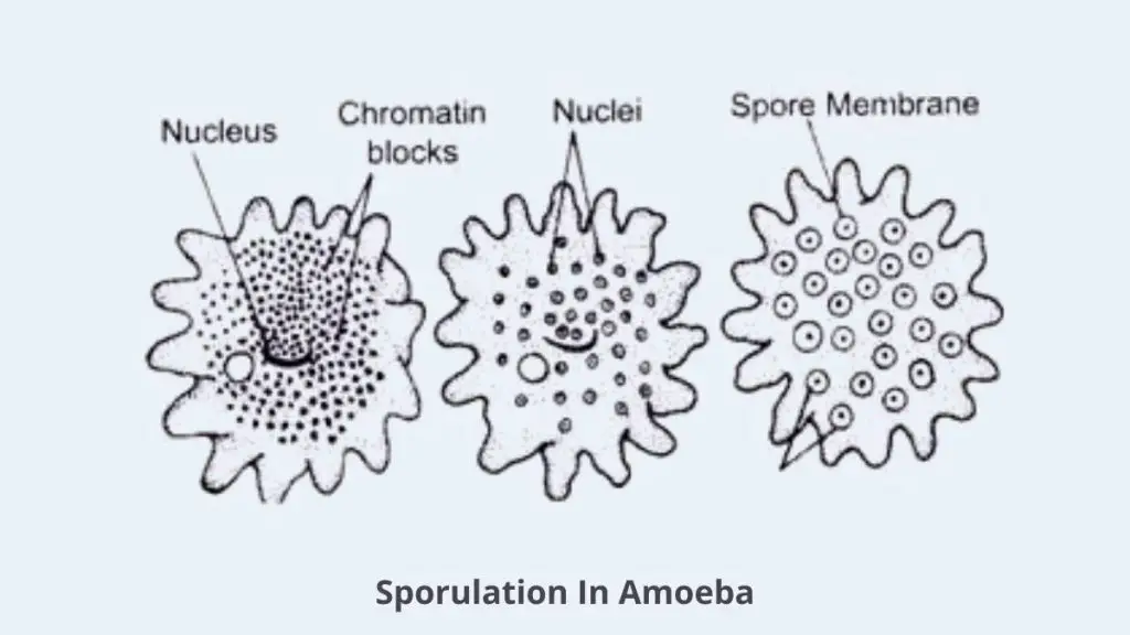
Draw well labelled diagram of different stages of binary fission in Amoeba and budding in yeast. Compare the features with established characteristics of both types of asexual reproduction in the given organism. Observations. Binary Fission in Amoeba

Amoeba Diagram Diagram Quizlet
The structure and diagram of amoeba. Amoeba. Paramecium Diagram. Body structure of an amoeba proteus. Amoeba. Amoeba. Irregular binary fission. Paramecium Dividing bacteria diagram. Amoeba is a single celled organism that appears transparent. Nutrition in an Amoeba occurs through a process called phagocytosis.

63 Amoeba Diagram Illustrations Clip Art Istock
Label Amoeba Anatomy Diagram Printout. EnchantedLearning.com is a user-supported site. As a bonus, site members have access to a banner-ad-free version of the site, with print-friendly pages.

In The Diagram Which Of The Following Processes Is Shown In Amoeba
Nutrition in an Amoeba occurs through a process called phagocytosis where the entire organism pretty much engulfs the food it plans on eating up. The mode of nutrition in amoeba is known as holozoic nutrition. It involves the ingestion, digestion and egestion of food material. Amoeba does not have any specialized organ for nutrition.

Amoeba Diagram Worksheet For 5th 6th Grade Lesson Planet

1 Simplest Way Of Drawing Amoeba How To Draw Amoeba In Easy Way Youtube Cute Henna Cute Henna Tattoos Science Diagrams
66 Amoeba Diagram Vector Images Amoeba Diagram Illustrations Depositphotos

Amoeba Proteus Morphology Locomotion And Reproduction Protozoa

63 Amoeba Diagram Illustrations Clip Art Istock
Amoeba

Amoeba Anatomy Vector Images And Illustration
Amoeba Structure Images Stock Photos Vectors Shutterstock
Feeding And Digestion In Amoeba Class 7 Science Lesson Nutrition In Animals

Amoeba Wikipedia
Amoeba Clipart Clip Art Library

66 Amoeba Diagram Vector Images Amoeba Diagram Illustrations Depositphotos

Diagram Of An Amoeba Stock Photos And Images Agefotostock
Amoeba Cell Characteristics Structure Movement Nutrition Reproduction Disease Habitat

63 Amoeba Diagram Illustrations Clip Art Istock

Describe The Feeding Habits Of Amoeba With Proper Diagram Brainly In
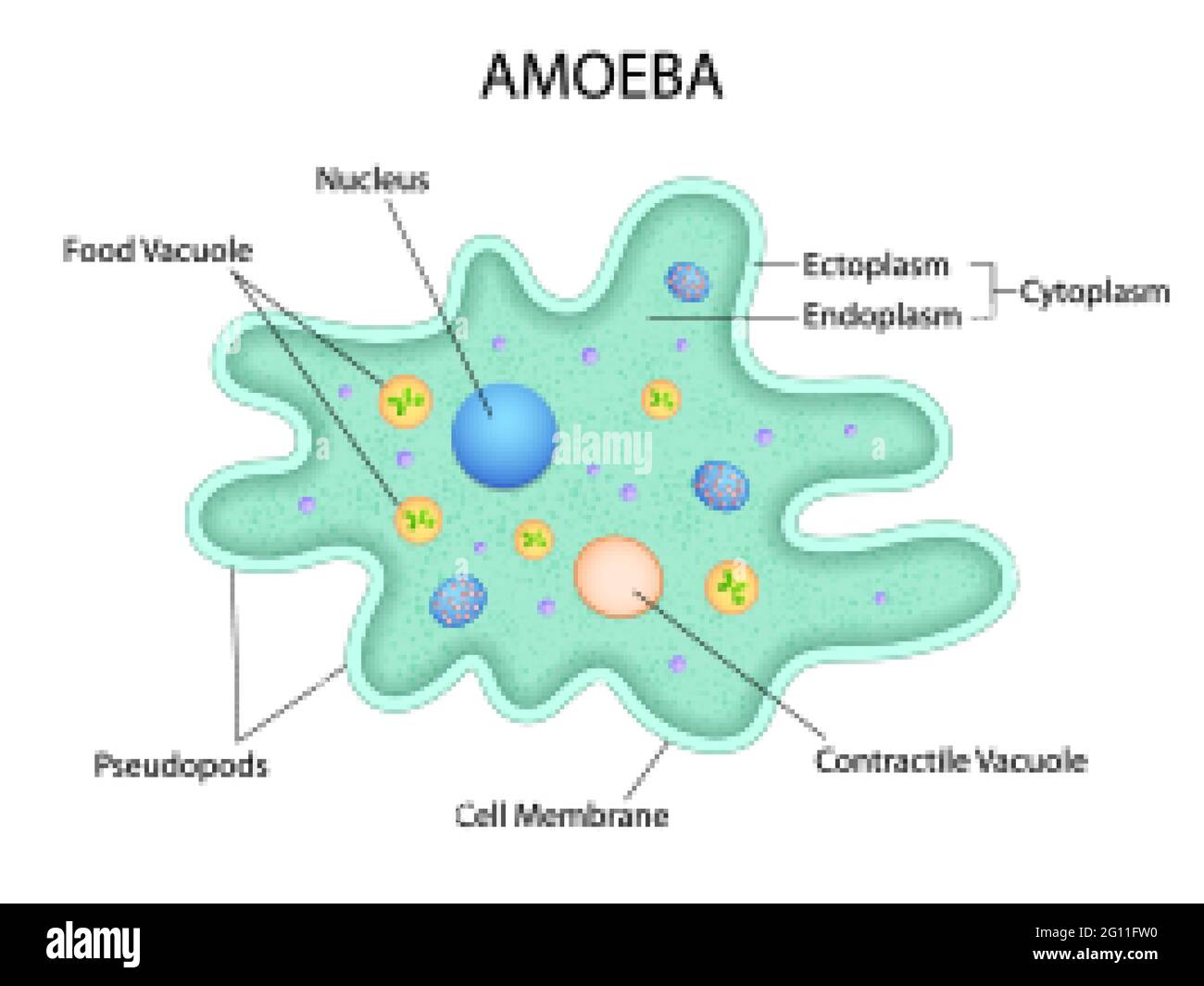
Illustration Of Healthcare And Medical Education Drawing Chart Of Amoeba For Science Biology Study Stock Vector Image Art Alamy

How To Draw Amoeba Amoeba Diagram Drawing Amoeba Drawing Amoeba Drawing Step By Step Video Youtube
Short Answer Question Draw A Diagram To Show The Nutrition In Amoeba And Label The Parts Used For This Purpose Mention Any Other Purpose Served By This Part Other Than Nutrition Snapsolve
35 Diagram Of Amoeba With Label Labels Database 2020

Science Ch 3 Amoeba Diagram Diagram Quizlet

Amoeba Proteus Characteristics Classification And Diagram Well Labelled
Comments
Post a Comment