39 rat heart diagram
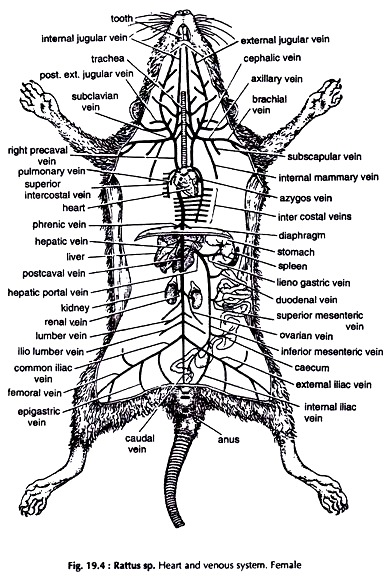
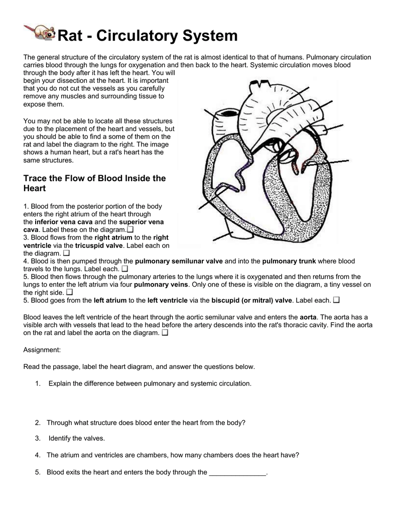
Veins (see diagram page 9) Your rat specimen has been double injected with latex to help you identify veins and arteries. Veins carry used blood (blue) back to the heart and lungs. The lungs re-oxygenate the blood and the heart pumps it back to the rest of the body. In the human body, these veins are not the same bright blue that you see in ... Secrets of the Four Chambers Revealed by Reptile Hearts. The molecular blueprint for evolution from cold-blooded to warm-blooded has been found. Embryo hearts show evolution of the heart from 3-chambered in frogs to 4-chambered in mammals. Watch an interview with developmental cardiologist Benoit Bruneau.
Download scientific diagram | Representative areas of rat heart showing (A) edema and (B) inflammatory cells. from publication: Treatment with nebivolol combined with physical training promotes ...

Rat heart diagram
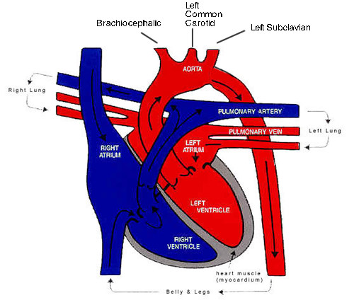
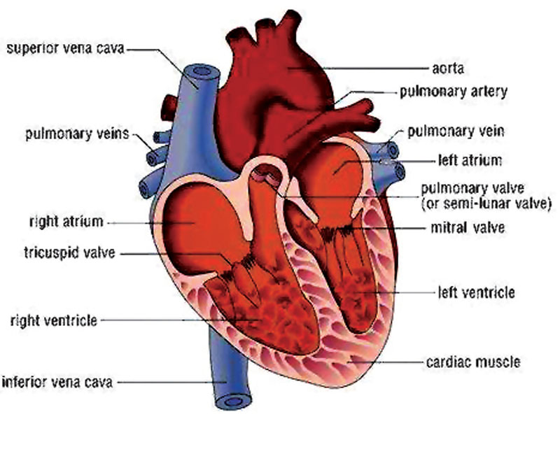
Circulatory system diagrams are visual representations of the circulatory system, also referred to as the cardiovascular system. It is comprised of three parts: the pulmonary circulation, coronary circulation, and systemic circulation. The main function of the circulatory system is to circulate blood, which carries oxygen and nutrients ... 2] cardiac muscle = striated; musculature of the heart 3] skeletal muscle = striated; generally attached to bone; usually under voluntary control Skeletal Muscle Skeletal (striated) muscle is composed of elongate, multinucleated cells (muscle fibers). Different types of muscle fibers are found among the various skeletal muscles of the body, e.g., The circulatory system and the anatomy of the heart of a rat are quite similar to that of humans. The heart of a rat acts like a pumping organ, while the blood vessels circulate the blood within the body and the lungs oxygenate the blood. Learn more about the circulatory system of a rat by going through this article.
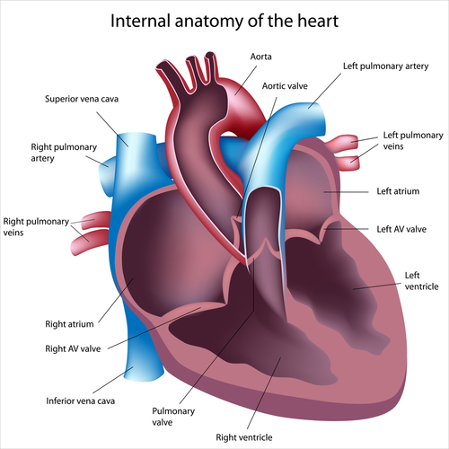
Rat heart diagram. There are 4 chambers, labeled 1-4 on the diagram below. To help simplify things, we can convert the heart into a square. We will then divide that square into 4 different boxes which will represent the 4 chambers of the heart. The boxes are numbered to correlate with the labeled chambers on the cartoon diagram. 5. Locate the teats on the ventral surface of the rat. Check a rat of another sex and determine whether both sexes have teats. 6. Examine the tail, the tails of rats do not have hair. (Some rodents, like gerbils, have hair on their tails.) 7. Locate the anus, which is ventral to the base of the tale. 8. ming instructions of rat and mouse protocol organs and tis sues in regulatory type toxicity studies. It is based on the experience made in the European RITA and American NACAD working groups and is an extended revision of trimming guides published in 1995 (B AHNEMANN et al.). The optimum localization for tissue preparation, the sample Anatomy of heart interior, frontal section. - stock illustration. Anatomy of heart interior, frontal section. : stock illustration. Buy the print. Get this image in a variety of framing options at Photos.com. Save to Board. Save to Board. New Board. SAME SERIES. View all.
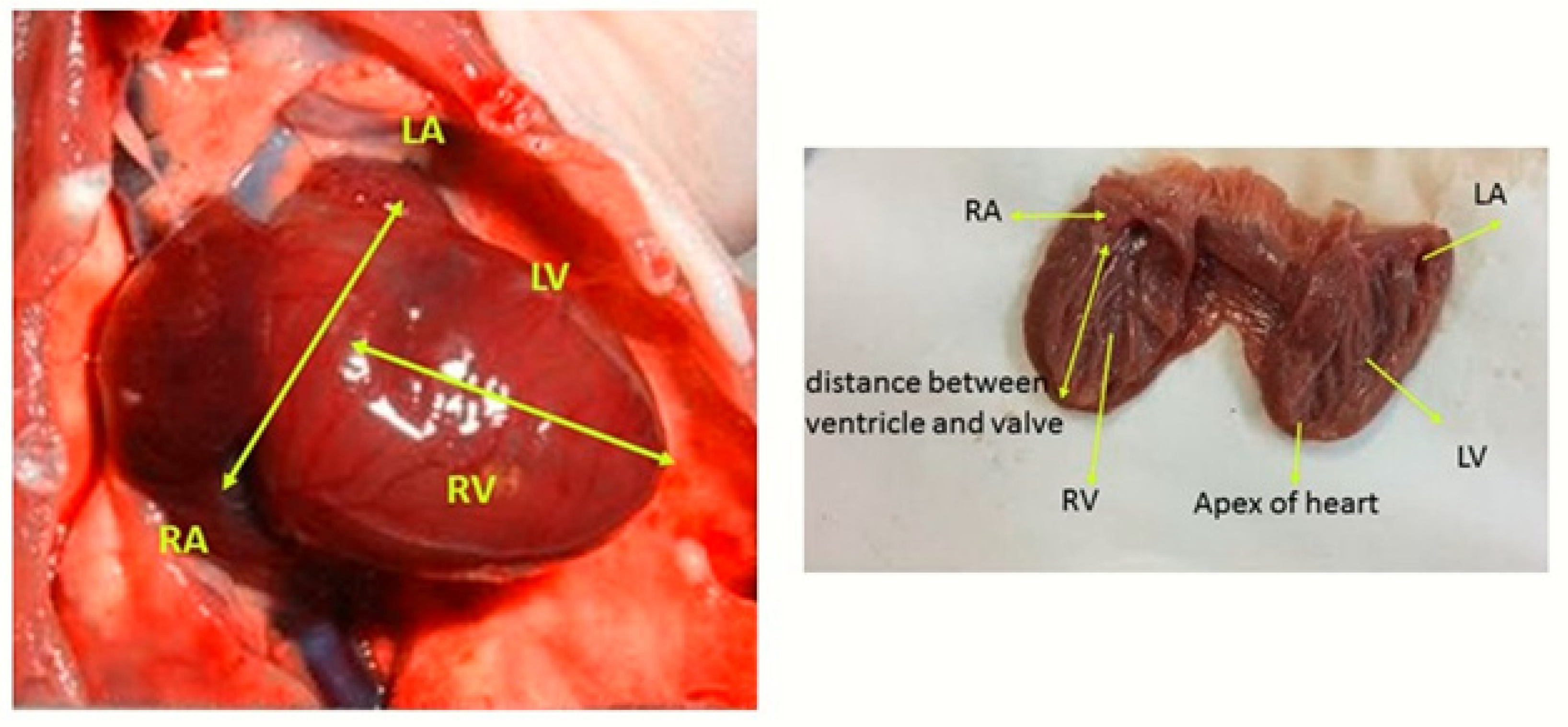
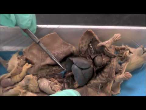
The heart of the rat is too small to view many of the structures listed below. Use models of human hearts or images in the dissection guides to identify the structures and label them on the diagram. 1. The heart is covered by a thin membrane called the pericardium. (We will come back to the heart later.) 3. Locate the thymus gland, which lies directly over the upper part of the heart. The thymus functions in the development of the immune system and is much larger in young rats than it is in older rats. 4. The rat is a typical mammal. Formerly guinea pig (Cavia sp.) (Fig. 19.1) were used for dissection in most of the undergraduate and postgraduate colleges in Indian Universities. Of late, due to prevailing high cost, guinea pig is being replaced by rat. Four species of rats are common in India, of which three are wild. 1. Observe the interior of the rat for any veins and arteries. Veins carry used blood (blue) back to the heart and lungs. Arteries carry oxygenated blood to the muscles and organs that need it. The arteries in your rat should be stained red for easy identification. Use Figures 3 and 4 to help you locate the major veins and arteries.
Rat Anatomy. Tap or move your mouse over the rat diagram below to view the organ names. *Tip: on smaller devices, you may want to flip your device to landscape if you can't see the full width of the image. Medical Illustration by Chris McKee for the Rat Guide. Additional descriptions will be added by the Rat Guide Team. Review of Rat Anatomy These pages will show you pictures of parts of a dissected rat with structures identified by numbers. To quiz yourself, see if you can identify the numbered parts. You can then check your answers by looking at another page which will show the same picture with all the parts labled. 502 Bella. March 27, 2006 at 16:08 (last edited February 10, 2021 at 14:18) The organs of the female reproductive system are specialized to produce ova (eggs), transport the egg cells to the site of fertilization, to provide a favorable environment for developing embryos, and to move offspring outside of the body (birth) at the appropriate time. The heart is located approximately at the level of the elbow. Place the needle, bevel up, into the chest, and puncture the heart. Apply slight back pressure with the syringe. If the needle is in the heart, blood will flow into the syringe. Wait until blood has filled the syringe before adding additional back pressure on the syringe.

Circulatory System Welcome To Rat Dissection
Download scientific diagram | Microphotograph of rat heart (H & E stain) (A) Cardiac representative section of various groups showing normal histology, (B) DMSO + Olive oil group, (C) CCl4 group ...
2
The surface projections of the heart represent points on the thoracic wall that map out the outline and valves of the heart. These include four borders (superior, right, inferior, left) and four valves (left atrioventricular, right atrioventricular, aortic, pulmonary).The main reference points used for the surface projections of the heart are the borders of the sternum and costal cartilages ...

The Bicuspid Valve In The Atrio Ventricular Opening Of The Left Half Of The Rat S Heart Atlas Of Animal Anatomy And Histology
Dec 15, 2012 - Continues the dissection of the rat by looking at the heart and circulatory system, including the major vessels such as the aorta, the femoral, and the subclavian.
Diagnostics Free Full Text Investigating Cardiac Morphological Alterations In A Pentylenetetrazol Kindling Model Of Epilepsy Html
This is how you can draw human heart in very easy way, stay tuned for more videos like this.
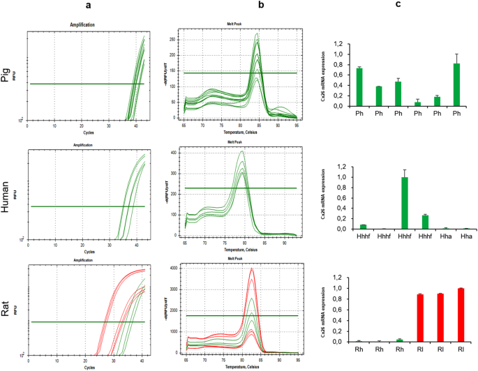
Connexin 26 Expression In Mammalian Cardiomyocytes Scientific Reports
(B) Venn diagram of identified backsplices and their conservation. (C) Density histogram of relative enrichment of backsplice-spanning reads from highly expressed rat circRNA candidates (detected with ≥ 3 reads in either all NRH or all ARH mock treated samples, n = 988) and regular splice junction spanning reads after RNase R digestion.
Rat Circulatory
• Rat: 20-25 gauge needle and 10-20 ml syringe • Anesthesia Technique: 1. Anesthetize animal. (Surgical plane of anesthesia is required!) 2. Test for reaction by corneal reflex and toe pinch. 3. Blood may be obtained through a ventral, left lateral, or open approach. 4. Ventral approach (closed) a. Place animal on back (dorsal recumbency) b.

Rat Reproduction Carolina Com
Your rat should be double injected so that arteries and veins are visible as blue and pink. Trace the Flow of Blood Inside the Heart. The heart of the rat is too small to view many of the structures listed below. Use models of human hearts or images in the dissection guides to identify the structures and label them on the diagram. 1.

Chronological And Morphological Study Of Heart Development In The Rat Marcela 2012 The Anatomical Record Wiley Online Library
Ultrasonographic evaluation of fetal development in the rat "The study objectives were to measure gestational sac (GS) diameter and crown-to-rump (CR) length in conscious pregnant rats and to determine the chronological ultrasonographic appearance of heart beat and fetal organogenesis. The study formed part of a unilateral surgical salpingectomy trial with 16 female Sprague-Dawley rats (Rattus ...

Glycogen Turnover In The Isolated Working Rat Heart Journal Of Biological Chemistry
The rats in group 1 were used as control (no additives); AX was used to treat the group 2 rats; the injection isoproterenol (ISO, isoprenaline hydrochloride, Sigma 15627, St. Louis, MO, USA) was used to induce heart failure (the rats in group 3), and rats in group 4 were treated with AX, and then two weeks later injected with ISO (twice).

A Modified Biventricular Working Heterotopic Rat Heart Transplant Model With Pressure Volume Loops Cardiac Function Analysis The Journal Of Heart And Lung Transplantation
The diagram below illustrates the muscles of the ventral surface of the rat. Be able to identify those ... Lungs - appear spongy and on either side of the heart. The right lung on the rat has four lobes and the left lung has three. Each lung is covered by a thin layer of tissue called the

Chronological And Morphological Study Of Heart Development In The Rat Marcela 2012 The Anatomical Record Wiley Online Library
Lung Diagram | lesson 1: Pulse of Life| lesson 2: Keeps on Pumpin' | lesson 3: Under Pressure | lesson 4: Sounds of the Heart | lesson 4a: Valves and Pumps | lesson 5: Lub Dub (valves) | lesson 5a: The Heart as a Pump | lesson 6: Go With the Flow | lesson 7: Lung Model | lesson 8: Ins and Outs of Respiration | lesson 9: Catch Your Breath | lesson 10: O 2 CO 2 Skit | lesson 11: X-Rays | lesson ...

Revised Guides For Organ Sampling And Trimming In Rats And Mice Heart
The circulatory system and the anatomy of the heart of a rat are quite similar to that of humans. The heart of a rat acts like a pumping organ, while the blood vessels circulate the blood within the body and the lungs oxygenate the blood. Learn more about the circulatory system of a rat by going through this article.
Rat Anatomy Dissection 네이버 블로그
2] cardiac muscle = striated; musculature of the heart 3] skeletal muscle = striated; generally attached to bone; usually under voluntary control Skeletal Muscle Skeletal (striated) muscle is composed of elongate, multinucleated cells (muscle fibers). Different types of muscle fibers are found among the various skeletal muscles of the body, e.g.,
Dissection Of Rat With Diagram Zoology
Circulatory system diagrams are visual representations of the circulatory system, also referred to as the cardiovascular system. It is comprised of three parts: the pulmonary circulation, coronary circulation, and systemic circulation. The main function of the circulatory system is to circulate blood, which carries oxygen and nutrients ...
Experimental Muscle Cell Patches May Hold Promise For Failing Hearts The Himalayan Times Nepal S No 1 English Daily Newspaper Nepal News Latest Politics Business World Sports Entertainment Travel Life Style News

Rat Heart Sectioning To Yield Posterior And Anterior Sections Of Left Download Scientific Diagram

18 Circulatory System Ideas Human Anatomy And Physiology Circulatory System Heart Anatomy

Cardiovascular Sciencedirect
Left Ventricular Mechanoenergetics In Excised Cross Circulated Rat Hearts Under Hypo Normo And Hyperthermic Conditions Scientific Reports

Chronological And Morphological Study Of Heart Development In The Rat Marcela 2012 The Anatomical Record Wiley Online Library

Effects Of Cardioplegia On Vascular Function And The No Reflow Phenomenon After Ischemia And Reperfusion Studies In The Isolated Blood Perfused Rat Heart The Journal Of Thoracic And Cardiovascular Surgery

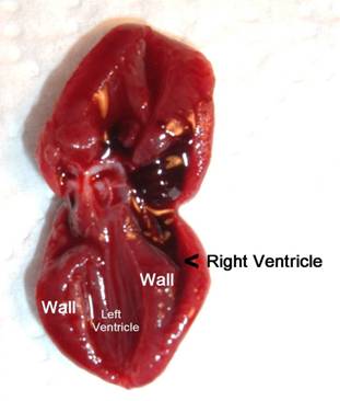
Chart No 172 Rat Heart
1
Rat Dissection Thoracic Cavity Circulatory System Youtube

Human Mustem Cell Grafting Into Infarcted Rat Heart Attenuates Adverse Tissue Remodeling And Preserves Cardiac Function Molecular Therapy Methods Clinical Development

Sutherlandandhearse

Reactivation Of Peroxisome Proliferator Activated Receptor A Is Associated With Contractile Dysfunction In Hypertrophied Rat Heart Journal Of Biological Chemistry

Heart And Sternum Revised Guides For Organ Sampling And Trimming In Rats And Mice
Click Read About The Rat Circulatory System Answer The Questions
Small Animal Pet Imaging Of Isolated Perfused Rat Heart Journal Of Nuclear Medicine

Cross Species Microrna Mrna Cardiac Atlas Data Sets Obtained For Rat Download Scientific Diagram

Rat Dissections
Comprehensive Analysis Of The Cardiac Proteome In A Rat Model Of Myocardial Ischemia Reperfusion Using A Tmt Based Quantitative Proteomic Strategy Proteome Science Full Text
Car Peptide Improves Remodulin Efficacy In Rat Model Of Ph
The Rat Report
1

Biology Honors Rat Lab Practical Thoracic Cavity Diagram Quizlet

Acylation Of Monolysocardiolipin In Rat Heart Journal Of Lipid Research

Predicting Left Ventricular Contractile Function Via Gaussian Process Emulation In Aortic Banded Rats Philosophical Transactions Of The Royal Society A Mathematical Physical And Engineering Sciences
Comments
Post a Comment