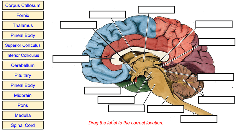
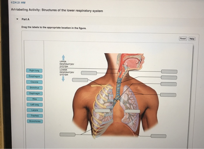
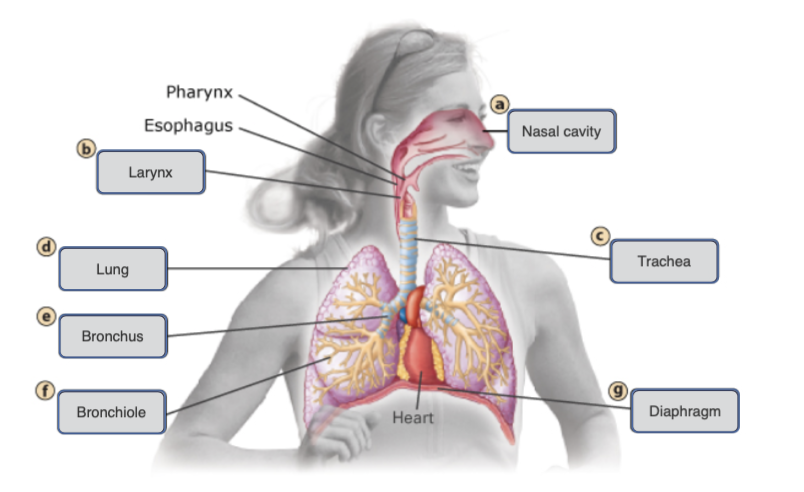
39 drag each label to the appropriate location on this diagram of the human respiratory system.
Anatomy and Physiology is a dynamic textbook for the yearlong Human Anatomy and Physiology course taught at most two- and four-year colleges and universities to students majoring in nursing and allied health. A&P is 29 chapters of pedagogically effective learning content, organized by body system, and written at an audience-appropriate level. Here is a Heart labeling quiz for you. The human heart is a vital organ for every human. The more healthy your heart is, the longer the chances you have of surviving, so you better take care of it. Take the following quiz to know how much you know about your heart. Questions and Answers. 1.
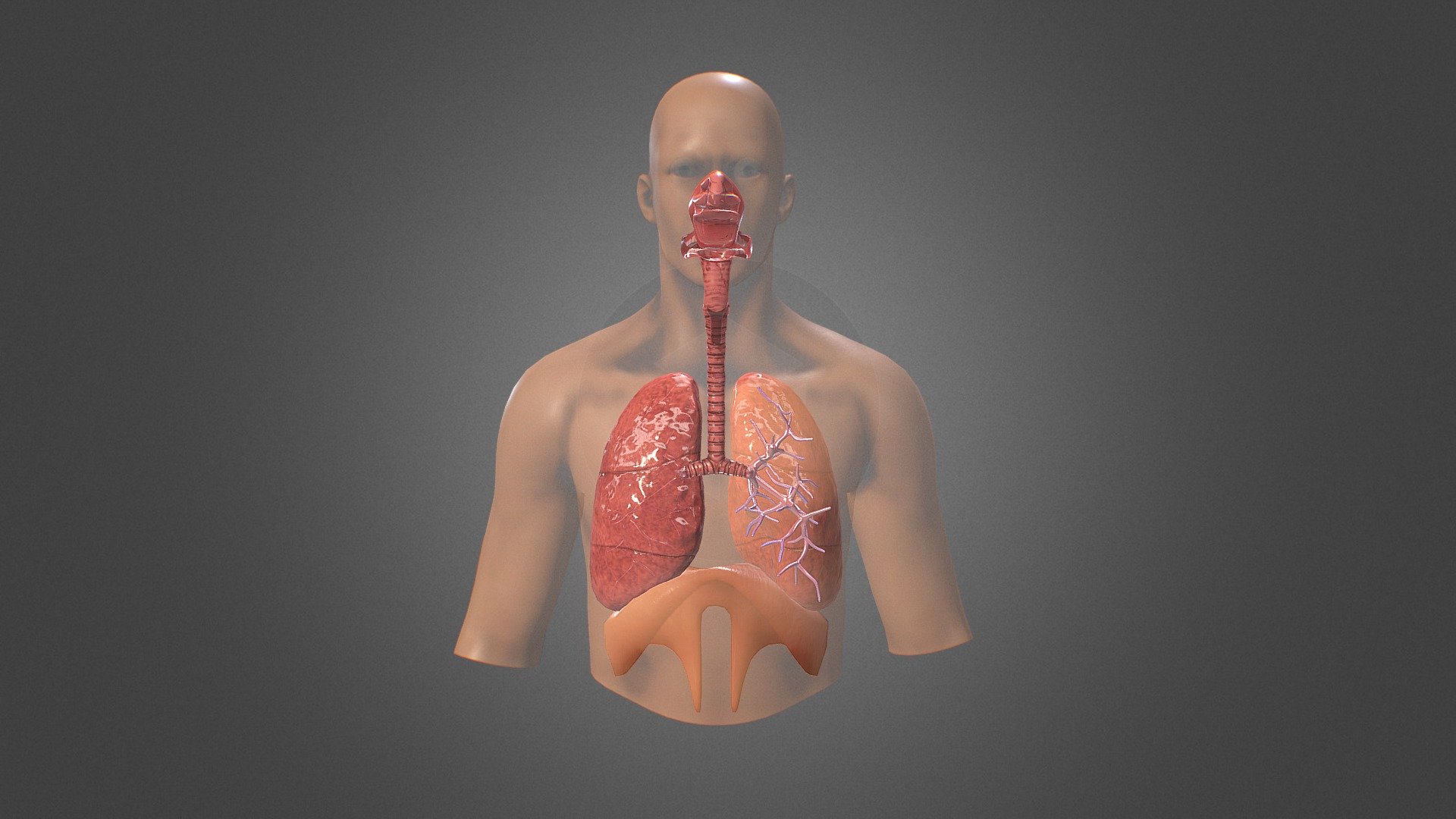
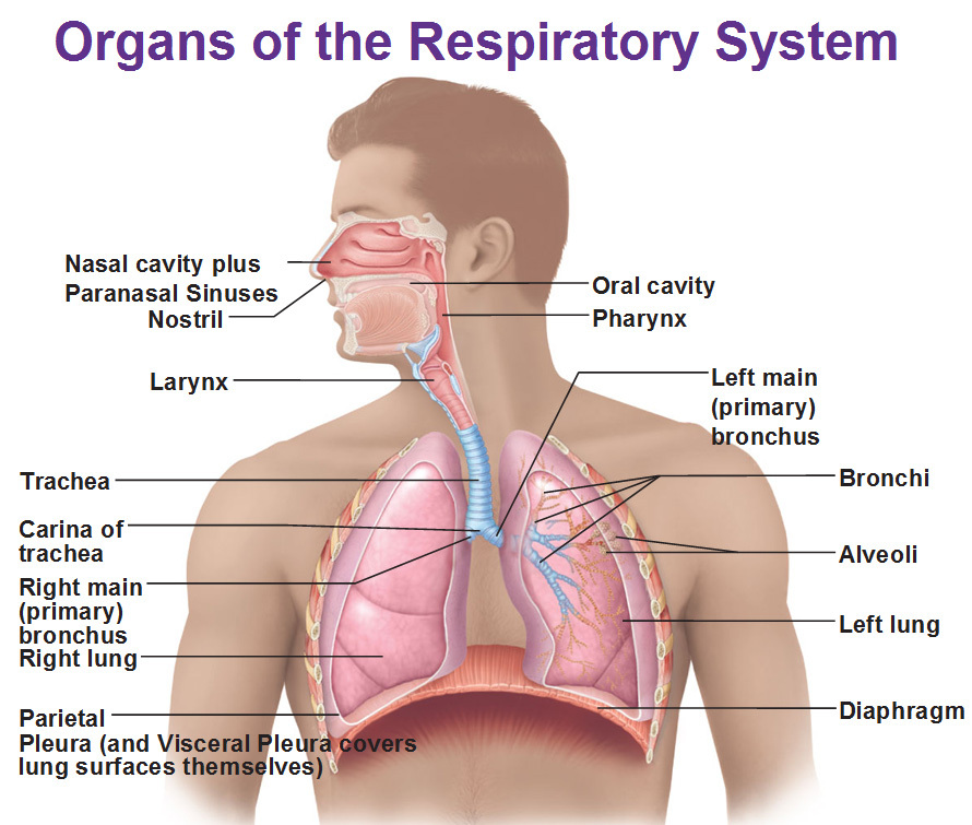
Diagram the relationship between multiple human body systems. 22 1 Organs And Structures Of The Respiratory System. Be sure to list the main functions and major organs of each body system explain how each system reacts to other systems identify a few medical conditions associated with each system and draw a picture of major organs of each system.
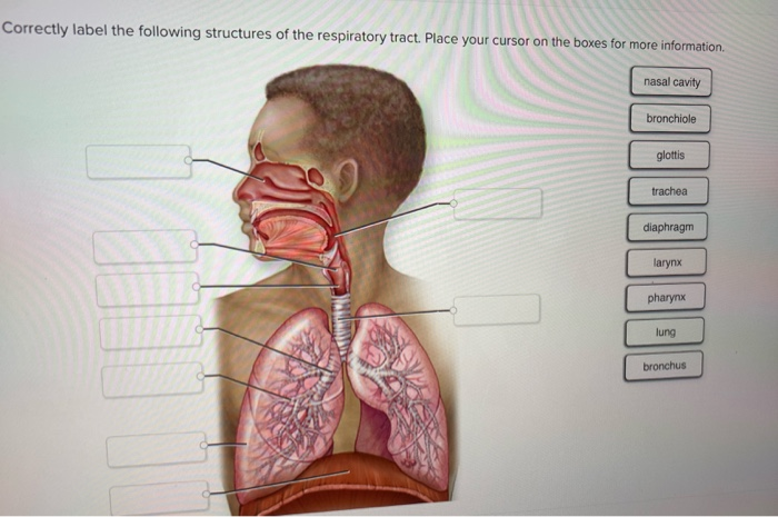
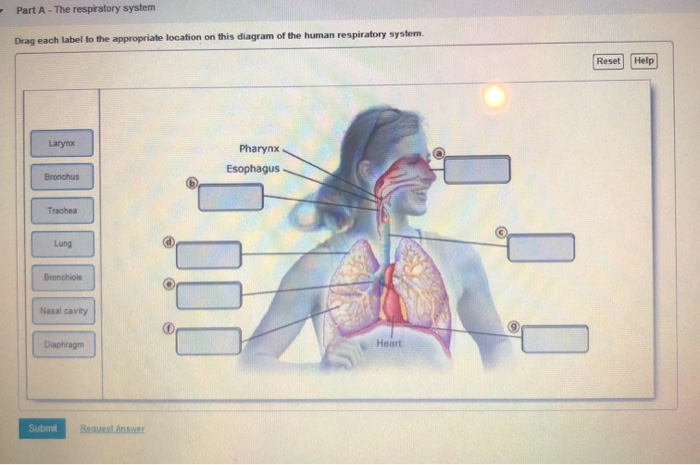
Drag each label to the appropriate location on this diagram of the human respiratory system.
Drag each label onto the appropriate figure, identifying whether the structure is associated with the large or small intestine. 32) Put the following layers of the digestive tract wall in order from the lumen to the deepest layer: 1. lamina propria 4. digestive (mucous) epithelium 2. muscularis externa 5. serosa Figure 4.8f Connective tissues. Figure: Diagram of parts of a microscope. There are three structural parts of the microscope i.e. head, base, and arm. Head - This is also known as the body, it carries the optical parts in the upper part of the microscope. Base - It acts as microscopes support. It also carries microscopic illuminators. The functions of the integumentary system are: Protection. The skin protects deeper tissues from mechanical damage (bumps), chemical damage (acids and bases), ultraviolet radiation (damaging effects of sunlight), bacterial damage, thermal damage (heat or cold), and desiccation (drying out). Temperature regulation.
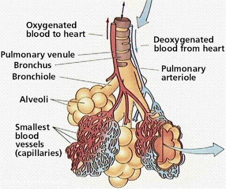
Drag each label to the appropriate location on this diagram of the human respiratory system.. Four Chambers of the Heart and Blood Circulation. The shape of the human heart is like an upside-down pear, weighing between 7-15 ounces, and is little larger than the size of the fist. It is located between the lungs, in the middle of the chest, behind and slightly to the left of the breast bone. The heart, one of the most significant organs ... Stroke volume: It refers to the quantity of blood pumped out of each ventricle with every heartbeat. Factors Determining Cardiac Output. The following factors determine the cardiac output of a human heart: Venous Return. This is the amount of blood that enters the heart through the veins per minute. Insulin is a peptide hormone produced by beta cells within the pancreas. It is responsible for regulating movement of glucose from the blood into cells. This article will consider the structure of insulin, how it is synthesised and secreted, its actions on the body and clinical conditions that are associated with faults in its production. Respiratory system diagram. Respiratory system organs, with the exception of the alveoli, function to conduct air into the lungs aided by the muscles of respiration (mainly the diaphragm and intercostal muscles). Once air is in the lungs it enters alveoli (the site of gas exchange) and interacts with blood transported by the pulmonary circulation.
Beyond mice, studies in non-human primates (NHPs) are instrumental in determining the efficacy of vaccine candidates for SARS-CoV-2, as the wild-type virus cannot efficiently replicate in common laboratory mouse strains, due to the lack of appropriate receptors to initiate viral infection . Briefly explain this comparison. Endocrine System WorksheetBiology 100C1. Label each number with the appropriate term. Use this simple Q. Endocrine system Short answer drag and drop worksheet on the endocrine system its organs and the hormones involved. Both the endocrine and nervous systems are major regulating systems of the body. 1222021 The ... The path of blood through the heart begins with the right atrium receiving blood, which has circulated through most of the body. This blood is relatively low in oxygen because most of it has already been delivered to different organs and tissues. This blood is also relatively high in carbon dioxide, which is a product of metabolism in the tissues. Cornea: It is the transparent, anterior or front part of our eye, which covers the pupil and the iris. The main function is to refract the light along with the lens. Iris: It is the pigmented, coloured portion of the eye, visible externally. The main function of the iris is to control the diameter of the pupil according to the light source.
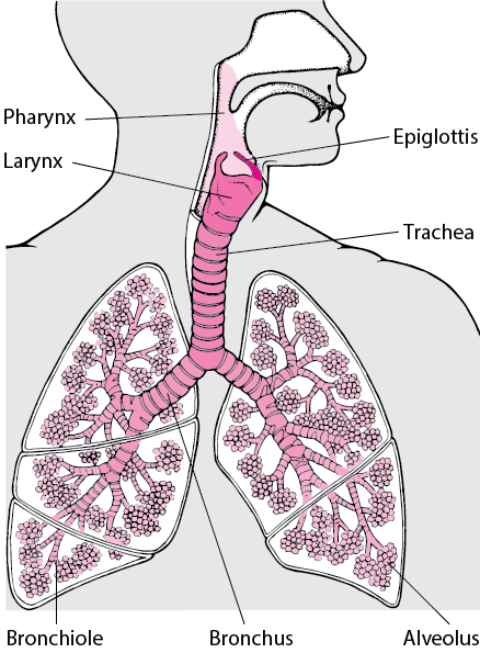
Author's Note: I made this as a kinda Wikipedia style biological summary of Valfalk/Valphalk/Valstrax with speculative features and explanations for its abilities. Do note that this is heavily speculative and mostly based on my very basic understanding of biology and general observation with these monsters. It is all labelled so you can skip to specific parts if you want. However, I put a lot of effort into this one in particular (Almost double the word count on the Zinogre post) and I’d recomme... When sunlight reflects off the near side, we call it a full Moon. The rest of the month we see parts of the daytime side of the Moon, or phases. These eight phases are, in order, new Moon, waxing crescent, first quarter, waxing gibbous, full Moon, waning gibbous, third quarter and waning crescent. The cycle repeats once a month (every 29.5 days). Respiratory System Diagram. Muscles of the Respiratory System. The diaphragm is a dome-shaped muscle that curves upwards towards the lungs. When it contracts, it becomes flattened and therefore increases the volume of the thoracic cavity. Similarly, contraction of the external intercostal muscles moves the ribs upwards and outwards. Corpus callosum: A band of white matter that joins the halves of the cerebrum at the deep center of the brain and coordinates nerve signals between each half. Cerebral arteries: Blood vessels that supply the cerebrum with oxygen-rich blood from the heart. There are three cerebral arteries: anterior (front), middle, and posterior (back).; Circle of Willis: A loop of cerebral arteries and other ...
Solved Correctly Label The Following Structures Of The Chegg Com
Integumentary system quiz and answers. One of the best ways to start learning about a new system, organ or region is with a labeled diagram showing you all of the main structures found within it. Not only will this introduce you to several new structures together, it will also give you an overview of the relations between them.

Drag The Labels Onto The Diagram To Identify The Structures Of The Upper Respiratory System Part Homeworklib
The Respiratory system: Educational information provided by the NIH. Includes an overview of the respiratory system, what happens when you breathe, what controls your breathing, and lung diseases and conditions. Inner body: Respiratory system: Includes 2D and 3D interactive respiratory system anatomy explorer. Provides information on each ...

8 504 Human Respiratory System Stock Vector Illustration And Royalty Free Human Respiratory System Clipart
WHMIS stands for the Workplace Hazardous Materials Information System. It is a comprehensive system for providing health and safety information on hazardous products intended for use, handling, or storage in Canadian workplaces. A WHMIS program ensures that the information about hazardous products is effectively communicated to workers.

5 1 Organs And Systems Of The Human Organism Medicine Libretexts
The anatomy of the heart is made easy in this post using labeled diagrams of the main cardiac structures and vascular system! We will review the anatomy, function, and order of blood through the human heart using pictures of the atria, ventricles, tricuspid valve, mitral valve, pulmonary valve, aortic valve, superior and inferior vena cava ...
Answered Drag The Labels To The Appropriate Bartleby
Michael Menna, DO, is a board-certified, active attending emergency medicine physician at White Plains Hospital in White Plains, New York. An organ system is a group of organs that work together to perform a complex function. There are eleven organ systems in the human body. All of these are ...
Answered Drag The Labels To The Appropriate Bartleby
The epidermis is the outermost layer of our skin. Its main purpose is protection. The epidermis is made of four main layers, or strata: The stratum corneum is the thick, tough outer layer. It is ...

Respiratory System Diagram Worksheets Teaching Resources Tpt
GRCh37(hg19) cytoband information was used to convert each CNV to a p- or q- arm-level change for simplification based on its location. Each CNV was annotated to be either a gain or a loss.

A P Lab Test 4 Flashcards Quizlet
The larynx is a cartilaginous segment of the respiratory tract located in the anterior aspect of the neck. The primary function of the larynx in humans and other vertebrates is to protect the lower respiratory tract from aspirating food into the trachea while breathing. It also contains the vocal cords and functions as a voice box for producing sounds, i.e., phonation.
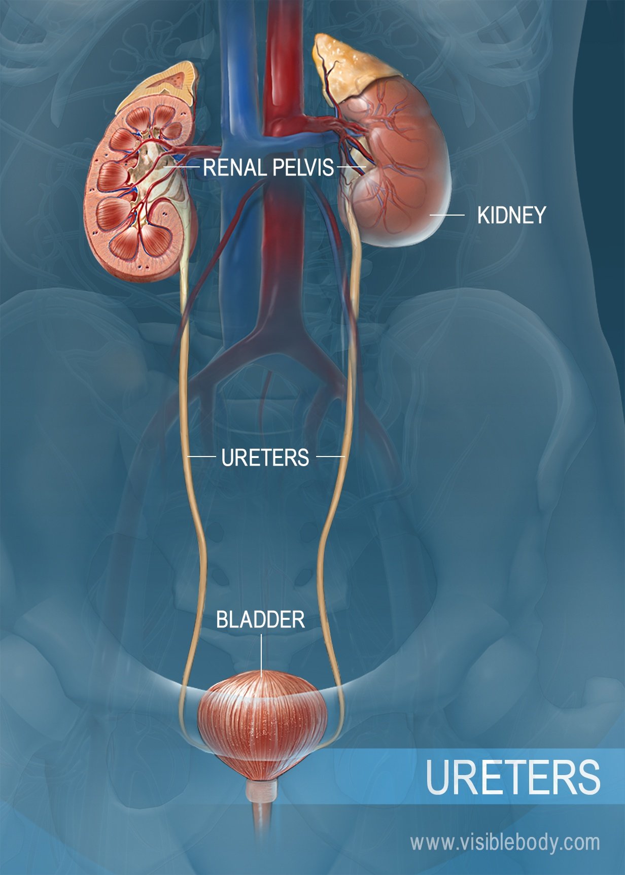
Urinary System Structures
Each one is approximately 2 to 3 centimeters wide and 7 to 8 centimeters long. The size of the labia majora is related to the amount of fat present. ... The Central Nervous System in the Human ...

Form 3 2 1 Human Respiratory System Biology Quizizz
Human body, the physical substance of the human organism. Characteristic of the vertebrate form, the human body has an internal skeleton with a backbone, and, as with the mammalian form, it has hair and mammary glands. Learn more about the composition, form, and physical adaptations of the human body.

Mcq Carefully Study The Diagram Of The Human Respiratory System
a run-away chemical reaction (chemical energy), the release of compressed gas or steam (pressure; high temperature), entanglement of hair or clothing in rotating equipment (kinetic energy), or. contact with electrodes of a battery or capacitor (electrical energy). Please see the OSH Answers on Hazard Identification for more information.
Brain Label Remote
Labeling, ranking, sorting, or sentence completion questions. All of these question types require you to position items into an area of the answer box. Answer these kinds of questions on a computer, not on a smartphone. Press Tab to move forward or Shift/Tab to move backwards through the provided answer items.
Human Respiratory System Review Buy Royalty Free 3d Model By Shinjou Shinjou 2509111
The functions of the integumentary system are: Protection. The skin protects deeper tissues from mechanical damage (bumps), chemical damage (acids and bases), ultraviolet radiation (damaging effects of sunlight), bacterial damage, thermal damage (heat or cold), and desiccation (drying out). Temperature regulation.

Sbi3u
Figure: Diagram of parts of a microscope. There are three structural parts of the microscope i.e. head, base, and arm. Head - This is also known as the body, it carries the optical parts in the upper part of the microscope. Base - It acts as microscopes support. It also carries microscopic illuminators.
Drag Each Label To The Appropriate Location On This Diagram Of The Human Respiratory System Wiring Site Resource
Drag each label onto the appropriate figure, identifying whether the structure is associated with the large or small intestine. 32) Put the following layers of the digestive tract wall in order from the lumen to the deepest layer: 1. lamina propria 4. digestive (mucous) epithelium 2. muscularis externa 5. serosa Figure 4.8f Connective tissues.
Overview Of The Respiratory System Lung And Airway Disorders Merck Manuals Consumer Version

Circulatory System Wikiwand

6 0wh9phwy3klm
Drag Each Label To The Appropriate Location On The

35 Drag Each Label To The Appropriate Location On The Flowchart Labels Design Ideas 2020

A P2 Ch22 Respiratory System Homework Flashcards Quizlet

Human Anatomy And Physiology I Chapter 21 Homework Flashcards Quizlet

Body Organization Ppt Download

A P Lab Test 4 Flashcards Quizlet

Click And Drag Each Label To The Appropriate Dock Chegg Com Plasma Membrane Membrane Structure Cell Membrane
Drag Each Label To The Appropriate Location On This Diagram Of The Human Respiratory System Wiring Site Resource
Solved Ch 21 Hw Art Labeling Activity Structures Of The Chegg Com
Right Ventricle Radiology Reference Article Radiopaedia Org

Sbi3u
Solved Part A The Respiratory System Drag Each Label To Chegg Com

Drag Each Label To The Correct Location On The Image Understand Payments Involved With Insurance Is Homeworklib

A P2 Ch22 Respiratory System Homework Flashcards Quizlet

Drag Each Label To The Correct Location On The Diagram Identify How A Hormone Molecule Performs Its Function

File Sinusrhythmlabels Svg Wikimedia Commons

A Hierarchical Structure For Representing 3d Respiration Organ Models Springerlink
2
Plants Free Full Text Protein Carbonylation Emerging Roles In Plant Redox Biology And Future Prospects Html
Respiratory System Flashcards Chegg Com
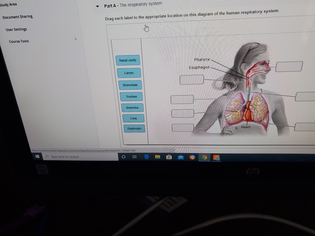
Solved Study Area Part A The Respiratory System Document Chegg Com
Comments
Post a Comment